 Back
BackBlood Vessels: Structure, Function, and Types
Study Guide - Smart Notes
 Tailored notes based on your materials, expanded with key definitions, examples, and context.
Blood Vessels
Overview of Blood Vessel Function
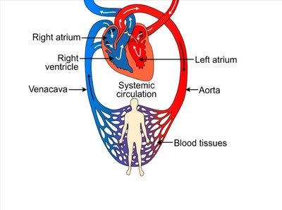
Blood vessels are essential components of the cardiovascular system, responsible for transporting blood throughout the body. They form a closed circuit that begins and ends at the heart, ensuring the delivery of oxygen, nutrients, and the removal of waste products from tissues.
Arteries: Carry blood away from the heart to the tissues.
Veins: Return blood from the tissues back to the heart.
Capillaries: Microscopic vessels where exchange of gases, nutrients, and wastes occurs between blood and tissues.
General Structure of Blood Vessels
Layers of Vessel Walls
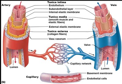
Most blood vessels (except capillaries) share a similar wall structure composed of three layers, or tunics:
Tunica intima: Innermost layer, consisting of endothelium (simple squamous epithelium) that lines the lumen of all vessels.
Tunica media: Middle layer, primarily smooth muscle and elastic fibers, responsible for vasoconstriction and vasodilation.
Tunica externa (adventitia): Outermost layer, composed of collagen fibers that protect and anchor the vessel.
Types of Blood Vessels
Arteries
Arteries are classified based on their size and function:
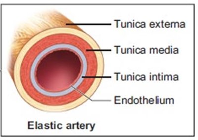
Elastic arteries: Largest arteries (e.g., aorta), rich in elastic fibers, act as pressure reservoirs to smooth out blood flow.
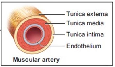
Muscular arteries: Medium-sized, contain more smooth muscle, distribute blood to specific organs.
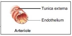
Arterioles: Smallest arteries, regulate blood flow into capillary beds through vasoconstriction and vasodilation.

Veins
Veins return blood to the heart and have thinner walls and larger lumens than arteries. They contain valves to prevent backflow, especially in the limbs.
Venules: Smallest veins, collect blood from capillaries.
Medium and large veins: Have all three tunics, but with a thinner tunica media and thicker tunica externa compared to arteries.
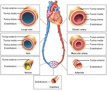
Capillaries
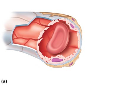
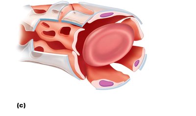
Capillaries are the smallest blood vessels, consisting only of endothelium and a basement membrane. They are the primary site for exchange between blood and tissues.
Continuous capillaries: Most common, least permeable, found in skin and muscle.
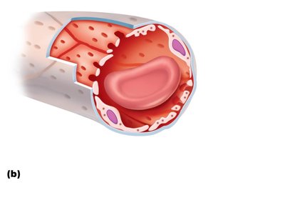
Fenestrated capillaries: Have pores (fenestrations) that increase permeability, found in kidneys and intestines.
Sinusoidal capillaries: Most permeable, with large gaps, found in liver, bone marrow, and spleen.
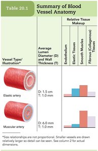
Summary Table: Blood Vessel Anatomy
The following tables summarize the structural differences among the major types of blood vessels:
Vessel Type | Average Lumen Diameter (D) | Wall Thickness (T) | Endothelium | Elastic Tissues | Smooth Muscle | Fibrous (Collagenous) Tissues |
|---|---|---|---|---|---|---|
Elastic artery | 1.5 cm | 1.0 mm | Present | High | Moderate | Moderate |
Muscular artery | 6.0 mm | 1.0 mm | Present | Low | High | Moderate |
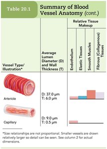
Arteriole | 37.0 μm | 6.0 μm | Present | Very low | Moderate | Low |
Capillary | 9.0 μm | 0.5 μm | Present | None | None | None |
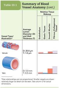
Venule | 20.0 μm | 1.0 μm | Present | None | Low | Low |
Vein | 5.0 mm | 0.5 mm | Present | None | Low | High |
Key Concepts and Applications
Pressure Gradient: Drives blood flow from high to low pressure regions.
Vasoconstriction (VC) and Vasodilation (VD): Adjust vessel diameter to regulate blood flow and pressure.
Valves in Veins: Prevent backflow, especially in limbs where blood must travel against gravity.
Capillary Exchange: Occurs via diffusion, intercellular clefts, vesicular transport, and fenestrations.
Example: The aorta (an elastic artery) expands as blood is ejected from the heart, then recoils to help maintain blood pressure during diastole.
Example: Capillaries in the kidneys are fenestrated to allow rapid filtration of blood plasma during urine formation.
Additional info: The structure of each vessel type is closely related to its function. For example, the thick tunica media in muscular arteries allows for precise control of blood flow to organs, while the thin walls of capillaries facilitate efficient exchange of materials.
