 Back
Bone Cells, Histology, and Ossification: Study Notes for ANP College Students
Study Guide - Smart Notes
 Tailored notes based on your materials, expanded with key definitions, examples, and context.
Chap 6 - part 2 - Bone Cells
Types of Bone Cells
Bone tissue is dynamic and constantly remodeled by the coordinated actions of several specialized cells. Understanding these cells is fundamental to grasping bone physiology and pathology.
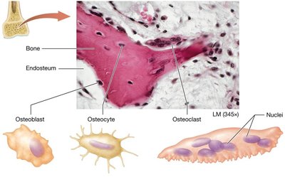
Osteoblasts: Cells responsible for bone formation. They secrete the organic matrix and initiate mineralization. Osteoblasts mature into osteocytes.
Osteocytes: Mature bone cells derived from osteoblasts. They maintain the bone matrix and communicate with other bone cells to regulate bone remodeling.
Osteoclasts: Large, multinucleated cells responsible for bone resorption. They break down bone matrix, releasing minerals into the blood.
Osteoblasts and Osteocytes
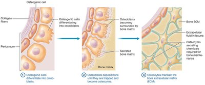
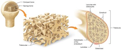
Osteoblasts are derived from osteogenic cells found in the inner periosteum and endosteum. They perform bone deposition by secreting the organic matrix (mainly collagen) and assisting in the formation of the inorganic matrix. Once osteoblasts become surrounded by the matrix, they differentiate into osteocytes and reside in small cavities called lacunae. Osteocytes maintain the extracellular matrix (ECM) and signal for bone remodeling in response to mechanical stress.
Osteoclasts
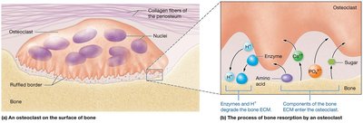
Osteoclasts are large, multinucleated cells with ruffled borders, derived from the fusion of cells formed in the bone marrow. They are responsible for bone resorption, a process in which they secrete hydrogen ions to dissolve the inorganic matrix and enzymes to break down the organic matrix. The breakdown products (minerals, amino acids, sugars) are released into the blood.
Clinical Connection: Osteopetrosis
Osteopetrosis ("Marble Bone Disease") is a condition where defective osteoclasts fail to resorb bone properly. This leads to increased bone mass, but the bone is weak and brittle. Severe forms can cause nerve entrapment, blindness, deafness, and reduced bone marrow space, which may be fatal in infants. Treatment may involve drugs to stimulate osteoclast activity and bone marrow function.
Histology of Bone
Compact Bone
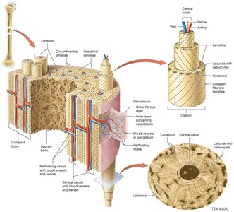
Compact bone forms the hard, dense outer shell of bones and is organized into structural units called osteons (Haversian systems). Each osteon consists of concentric rings called lamellae, with collagen fibers in alternating directions to resist twisting forces. The central (Haversian) canal contains blood vessels and nerves. Lacunae are small cavities between lamellae that house osteocytes. Canaliculi are tiny canals connecting lacunae, allowing communication between osteocytes. Osteons are connected by interstitial lamellae and surrounded by circumferential lamellae. Perforating (Volkmann) canals connect blood vessels between osteons and the periosteum.
Spongy Bone
Spongy bone (cancellous bone) is found inside bones and consists of a network of trabeculae. These trabeculae are covered with endosteum and contain concentric lamellae with lacunae and canaliculi, but lack central and perforating canals. Spongy bone provides structural support and houses bone marrow.
Bone Formation: Ossification
Overview of Ossification
Ossification (osteogenesis) is the process of bone formation, which continues through childhood. There are two main types of bone tissue formed during ossification:
Primary (woven) bone: Immature bone with irregular collagen bundles and abundant osteocytes. It is later replaced by secondary bone.
Secondary (lamellar) bone: Mature bone with organized lamellae and parallel collagen bundles, providing greater strength.
Types of Ossification
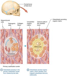
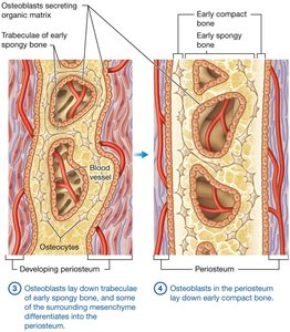
Intramembranous Ossification: Bone develops from a mesenchymal membrane. This process forms many flat bones, including most skull bones and the clavicles. The inner spongy bone forms first.
Endochondral Ossification: Bone develops from a hyaline cartilage model. This process forms most bones below the head (except the clavicles), with compact bone forming first.
Steps of Intramembranous Ossification
Intramembranous ossification involves the following steps:
Osteoblasts develop in the primary ossification center from mesenchymal cells.
Osteoblasts secrete organic matrix, which calcifies; trapped osteoblasts become osteocytes.
Osteoblasts lay down trabeculae of early spongy bone, and some mesenchyme differentiates into periosteum.
Osteoblasts in the periosteum lay down early compact bone. Larger bones require fusion of multiple ossification centers; fontanels in newborns are areas of incomplete fusion.
Endochondral Ossification
Endochondral ossification is the process by which most bones are formed, beginning during the fetal period. The hyaline cartilage model contains chondrocytes and cartilage ECM, surrounded by a perichondrium. Ossification begins at primary ossification centers, with long bones also developing secondary ossification centers in their epiphyses.
Chondroblasts in the perichondrium differentiate into osteoblasts, triggered by chemical signals.
Bone begins to ossify from the outside: osteoblasts build a bone collar on the external surface, while internal cartilage calcifies and chondrocytes die.
Study Strategies
Elaborative Questioning
To deepen understanding, use elaborative questioning. For example, ask "What are the four types of bone cells?" and then "How are they different?" Continue generating and answering related questions to reinforce learning and integration of concepts.
Summary Table: Bone Cell Types and Functions
Cell Type | Origin | Main Function | Location |
|---|---|---|---|
Osteogenic Cell | Mesenchyme | Stem cell for osteoblasts | Periosteum, endosteum |
Osteoblast | Osteogenic cell | Bone formation (deposition) | Periosteum, endosteum |
Osteocyte | Osteoblast | Maintains bone matrix | Lacunae in bone matrix |
Osteoclast | Fusion of bone marrow cells | Bone resorption | Bone surfaces |
