 Back
Bone Development, Growth, and Disorders: Study Notes for ANP College Students
Study Guide - Smart Notes
 Tailored notes based on your materials, expanded with key definitions, examples, and context.
CHAP 6 Part 3 - Endochondral Ossification
Overview of Endochondral Ossification
Endochondral ossification is the process by which most bones, especially long and short bones, are formed from a hyaline cartilage model. This process is essential for the development of the fetal skeleton and the growth of bones during childhood and adolescence.
Chondroblasts in the perichondrium differentiate into osteoblasts.
Osteoblasts build a bone collar on the bone’s external surface, initiating ossification from the outside.
Internal cartilage begins to calcify, and chondrocytes die due to lack of nutrients.
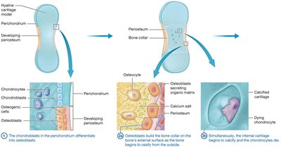
In the primary ossification center, osteoblasts replace calcified cartilage with early spongy bone.
Secondary ossification centers and the medullary cavity develop in the epiphyses.
As the medullary cavity enlarges, remaining cartilage is replaced by bone, and the epiphyses finish ossifying.
Cartilage remains in the epiphyseal plates (growth plates) until bone growth in length ceases, and as articular cartilage for life.
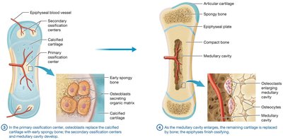
Example: The long bones of the limbs, such as the femur and humerus, develop through endochondral ossification.
Persistence of Hyaline Cartilage
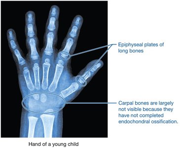
Hyaline cartilage persists in the epiphyseal plates of long bones and in the carpal bones of the hand during childhood, allowing for continued bone growth.
Comparison: Intramembranous vs. Endochondral Ossification
Key Differences
Types of Bones: Flat bones (e.g., skull bones) form by intramembranous ossification within a fibrous membrane. Long and short bones form by endochondral ossification from a cartilage model.
Order of Bone Formation: In endochondral ossification, compact bone forms first, followed by spongy bone. In intramembranous ossification, spongy bone forms first, and compact bone forms later.
Additional info: Intramembranous ossification is crucial for the development of the flat bones of the skull and the clavicle.
Bone Disorders
Osteoporosis
Osteoporosis is a bone disease characterized by inadequate inorganic matrix in the extracellular matrix (ECM), making bones brittle and increasing the risk of fractures.
Common causes include dietary deficiencies (calcium, vitamin D), female sex, advanced age, lack of exercise, hormonal changes (e.g., postmenopausal estrogen loss), genetic factors, and certain diseases.
Prevention and treatment involve adequate calcium and vitamin D intake, weight-bearing exercise, hormone replacement, and medications that affect osteoclast or osteoblast activity.

Achondroplasia
Achondroplasia is the most common cause of dwarfism, resulting from a genetic mutation affecting growth factor receptors on cartilage. This leads to decreased endochondral ossification and cell division in the epiphyseal plate, resulting in shortened limbs and characteristic features.
Symptoms include short stature, long trunk, abnormal spinal curvatures, and possible chest wall deformities.
Management may involve medications and surgeries.
Gigantism and Acromegaly
Both conditions are caused by excess secretion of growth hormone, but at different life stages:
Gigantism: Occurs before epiphyseal plate closure, causing excessive longitudinal and appositional bone growth.
Acromegaly: Occurs after epiphyseal plate closure, resulting in enlarged bones of the skull, face, hands, feet, and soft tissues.
Treatment typically involves removal of the hormone-secreting tumor.

Bone Growth in Length
Epiphyseal Plate Structure and Zones
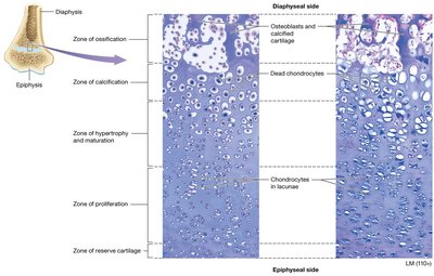
Longitudinal growth occurs at the epiphyseal plate, which consists of five distinct zones:
Zone of Reserve Cartilage: Inactive chondrocytes that can divide if needed.
Zone of Proliferation: Actively dividing chondrocytes in lacunae; main site of mitosis.
Zone of Hypertrophy and Maturation: Contains mature, enlarging chondrocytes.
Zone of Calcification: Contains dead, calcified chondrocytes; far from blood supply.
Zone of Ossification: Calcified cartilage is replaced by bone as osteoblasts invade.
Steps of Longitudinal Growth
Chondrocytes divide in the zone of proliferation.
Chondrocytes enlarge and mature in the next zone.
Chondrocytes die and their matrix calcifies.
Calcified cartilage is replaced with bone by osteoblasts in the zone of ossification.
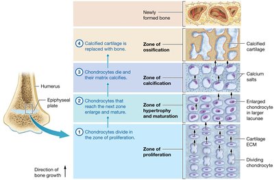
Longitudinal growth continues as long as mitosis occurs in the zone of proliferation. When ossification overtakes proliferation (typically between ages 13–21), the epiphyseal plate closes, leaving the epiphyseal line.
Bone Growth in Width (Appositional Growth)
Mechanism of Appositional Growth
Appositional growth increases the width of bones and can continue after longitudinal growth ceases. Osteoblasts between the periosteum and bone surface lay down new circumferential lamellae, thickening the compact bone of the diaphysis. Osteoclasts in the medullary cavity resorb inner lamellae, enlarging the cavity as the bone thickens.
The Role of Hormones in Bone Growth
Hormonal Regulation
Growth Hormone: Produced by the anterior pituitary gland; increases mitosis of chondrocytes, stimulates osteogenic cells, and triggers appositional growth.
Testosterone: Increases appositional growth and mitosis at the epiphyseal plate, leading to thicker bones and earlier closure of growth plates in males (by age 16–17).
Estrogen: Similar effects as testosterone but less pronounced; epiphyseal plates close earlier in females (by age 14–15).
Bone Remodeling
Definition and Importance
Bone remodeling is the continual process of bone formation (deposition) and bone loss (resorption). It serves several functions:
Maintaining calcium ion homeostasis
Bone repair
Replacement of primary bone with secondary bone
Replacement of older, brittle bone with newer bone
Adaptation of bone to tension and stresses
