 Back
BackBone Formation, Growth, Remodeling, and Fracture Repair
Study Guide - Smart Notes
 Tailored notes based on your materials, expanded with key definitions, examples, and context.
Bone Formation (Ossification)
Types of Ossification
Ossification is the process by which bone tissue is formed. There are two primary types of ossification: intramembranous ossification and endochondral ossification.
Intramembranous Ossification: Occurs within mesenchymal (embryonic connective) tissue and is responsible for forming flat bones such as those of the skull and mandible. This process does not involve a cartilage model; instead, mesenchymal cells differentiate directly into osteoblasts, which secrete bone matrix. The matrix then ossifies, forming trabeculae and eventually compact bone and periosteum.
Endochondral Ossification: Forms most long bones of the body. This process begins with a hyaline cartilage model, which is gradually replaced by bone tissue. The perichondrium (covering the cartilage) becomes the periosteum (covering the bone). Ossification centers develop in the diaphysis (primary) and epiphyses (secondary), leading to the formation of the medullary cavity, articular cartilage, and epiphyseal plate.
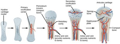
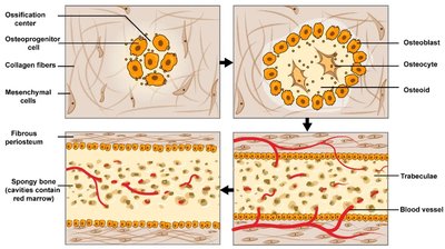
Key Steps in Endochondral Ossification:
Development and growth of the hyaline cartilage model
Formation of the bony collar and primary ossification center in the diaphysis
Development of secondary ossification centers in the epiphyses and formation of the medullary cavity
Formation of articular cartilage and the epiphyseal plate
Key Steps in Intramembranous Ossification:
Mesenchymal cells differentiate into osteogenic cells and osteoblasts
Osteoblasts secrete matrix, which ossifies
Osteoblasts become osteocytes
Trabeculae form, followed by periosteum and compact bone
Bone Growth
Growth in Length (Longitudinal Growth)
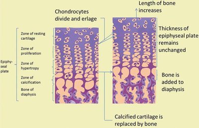
Bone lengthening occurs at the epiphyseal plates through a process involving the proliferation and maturation of cartilage, followed by its replacement with bone tissue. This process continues until the epiphyseal plate ossifies and becomes the epiphyseal line, marking the end of longitudinal growth.
Four zones in the epiphyseal plate: resting, proliferating, hypertrophic, and calcified cartilage
Chondrocytes divide and enlarge, increasing bone length
Calcified cartilage is replaced by bone tissue
Key Terms:
Proliferation: Increase in cell number
Hypertrophy: Increase in cell size
Calcification: Deposition of calcium salts; calcified cartilage is functionally dead
Growth in Width (Appositional Growth)
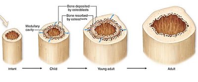
Bones increase in diameter through appositional growth, which involves the addition of new bone tissue by osteoblasts in the periosteum and the resorption of bone by osteoclasts in the endosteum. This process enlarges the medullary cavity and thickens the bone.
Osteogenic cells in the periosteum become osteoblasts, forming new matrix
Osteoclasts in the endosteum break down bone tissue, increasing the medullary cavity diameter
Bone Remodeling and Calcium Homeostasis
Bone Remodeling
Bone is a dynamic tissue that is constantly being resorbed by osteoclasts and deposited by osteoblasts. This process is essential for bone health, adaptation to stress, and repair of microdamage.
Requires minerals (Ca2+, P), vitamins (C, K, B12, A), and hormones (IGFs, T3, T4, estrogen, androgens, insulin)
Calcium Homeostasis
Calcium is stored in bone and is vital for muscle contraction, nerve impulse conduction, and protein function. Blood calcium levels are tightly regulated:
Normal range: 8–10 mg/dL
Low Ca2+: Parathyroid hormone (PTH) is released, stimulating osteoclasts to release calcium from bone
High Ca2+: Calcitonin is released, stimulating osteoblasts to deposit calcium into bone
Aging and Bone Tissue
Aging affects bone tissue in two main ways, both increasing the risk of fractures:
Demineralization: Loss of bone minerals, which may result in osteoporosis
Decreased protein (collagen) synthesis: Reduces bone flexibility and strength
Prevention strategies include nutritional supplements, exercise, and hormone therapy.
Bone Fractures and Repair
Types of Bone Fractures
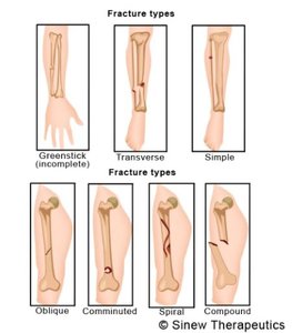
A fracture is any break in a bone. Common types include:
Simple (closed): Bone breaks but does not penetrate the skin
Compound (open): Broken ends protrude through the skin
Stress fracture: Microscopic fissures in bone
Comminuted: Bone is splintered or crushed
Greenstick: One side bends, the other side breaks (common in children)
Other types: Compression, spiral, oblique, impacted, transverse
Fracture Repair
Bone repair is a four-step process that restores both structure and function:
Fracture hematoma formation: Blood clot forms at the site of the break
Cartilaginous callus formation: Phagocytic cells clean debris; fibrocartilage forms a temporary bridge
Bony callus formation: Osteoblasts replace cartilage with bone; strength is restored (4–8 weeks after injury)
Remodeling: Excess bone is removed, and new osteons form to restore normal bone structure
Fracture Type | Description |
|---|---|
Simple (Closed) | Bone breaks but does not penetrate the skin |
Compound (Open) | Broken ends protrude through the skin |
Stress | Microscopic fissures in bone |
Comminuted | Bone is splintered or crushed |
Greenstick | One side bends, other side breaks |
Oblique | Diagonal break across the bone |
Spiral | Twisting force causes a spiral-shaped break |
Transverse | Break occurs at a right angle to the bone's axis |
Impacted | One broken end is driven into the other |
Additional info: Proper bone healing requires adequate blood supply, nutrients, and sometimes medical intervention (e.g., immobilization or surgery).
