 Back
BackBone Growth, Ossification, and Maintenance: A&P Study Guide
Study Guide - Smart Notes
 Tailored notes based on your materials, expanded with key definitions, examples, and context.
Bones and Bone Structure
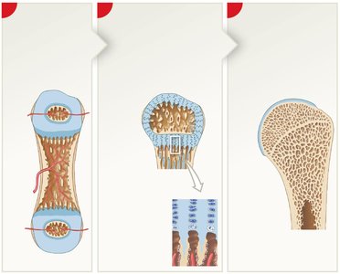
Endochondral Ossification
Endochondral ossification is the process by which most bones in the body are formed from a cartilage model. This multi-step process transforms hyaline cartilage into mature bone tissue, allowing for growth in both length and diameter.
Step 1: Chondrocytes in the center of the cartilage shaft enlarge, and the matrix calcifies. The chondrocytes die, leaving cavities.
Step 2: Blood vessels grow around the cartilage, and perichondrium cells become osteoblasts, forming a bone collar.
Step 3: Blood vessels penetrate the cartilage, fibroblasts become osteoblasts, and spongy bone forms at the primary ossification center.
Step 4: Remodeling creates a medullary cavity, and bone replaces cartilage near the epiphyses.
Step 5: Capillaries and osteoblasts migrate into the epiphyses, forming secondary ossification centers.
Step 6: The epiphyseal plate separates the epiphysis from the diaphysis, allowing continued growth.
Step 7: At puberty, epiphyseal cartilage production slows, osteoblast activity increases, and the epiphyseal plate closes, leaving an epiphyseal line.
Key Terms: Epiphysis, Diaphysis, Metaphysis, Epiphyseal plate, Osteoblast, Osteocyte, Spongy bone, Compact bone
Bone Growth at Epiphyseal Cartilages
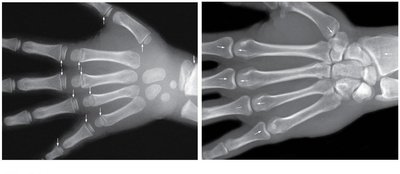
Bone growth in length occurs at the epiphyseal cartilages (growth plates). New cartilage is produced on the epiphyseal side, while osteoblasts replace cartilage with bone on the diaphyseal side. This process continues until the epiphyseal plate closes at adulthood.
Juvenile Skeleton: Visible epiphyseal cartilage allows for continued bone growth.
Adult Skeleton: Epiphyseal lines mark the former location of the growth plates, indicating growth has ceased.
Intramembranous Ossification
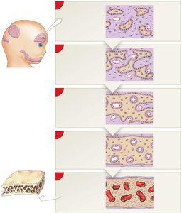
Intramembranous ossification forms flat bones such as those of the skull, mandible, and clavicle. It begins with mesenchymal cells differentiating into osteoblasts, which secrete osteoid and initiate bone formation directly within connective tissue.
Step 1: Mesenchymal cells cluster and differentiate into osteoblasts, secreting osteoid.
Step 2: Osteoblasts trapped in matrix become osteocytes; bone grows outward in spicules.
Step 3: Blood vessels grow between spicules, accelerating bone growth.
Step 4: Spongy bone forms, trapping blood vessels.
Step 5: Remodeling produces compact bone and periosteum.
Blood Supply to Mature Bone
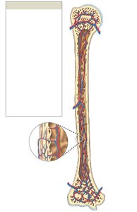
Mature bones receive blood through several sources, ensuring nutrient delivery and waste removal. The main vessels include nutrient arteries and veins, metaphyseal and epiphyseal arteries and veins, and periosteal vessels.
Nutrient Artery and Vein: Enter through the nutrient foramen to supply the diaphysis.
Metaphyseal and Epiphyseal Vessels: Supply the ends of the bone.
Periosteal Vessels: Supply the outer compact bone.
Hormonal Regulation of Bone Growth and Maintenance
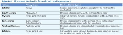
Bone growth and maintenance are regulated by several hormones, each with specific effects on the skeletal system. These hormones influence osteoblast and osteoclast activity, calcium absorption, and bone matrix synthesis.
Hormone | Primary Source | Effect on Skeletal System |
|---|---|---|
Calcitriol | Kidneys | Increases calcium and phosphate absorption by intestines |
Growth Hormone | Pituitary gland | Stimulates osteoblast activity and bone matrix synthesis |
Thyroxine | Thyroid gland | Stimulates osteoblast activity and bone matrix synthesis |
Sex Hormones | Ovaries/Testes | Stimulate osteoblast activity; estrogen causes faster epiphyseal closure |
Parathyroid Hormone | Parathyroid glands | Stimulates osteoclast activity; increases blood calcium ion level |
Calcitonin | Thyroid gland (C cells) | Decreases blood calcium ion level; inhibits osteoclasts |
Disorders of Bone Growth
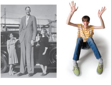
Abnormalities in bone growth can result from genetic or hormonal imbalances. Two notable disorders are gigantism and Marfan syndrome.
Gigantism: Excess growth hormone causes abnormal bone growth and increased height.
Marfan Syndrome: Genetic disorder affecting connective tissue, leading to long limbs and fingers.
Chemical Composition of Bone
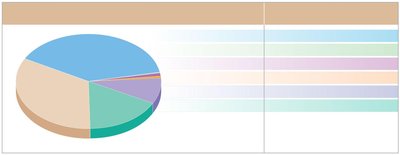
Bones are composed of both organic and inorganic components. The organic matrix (mainly collagen) provides flexibility, while inorganic minerals (primarily hydroxyapatite) give strength and rigidity.
Organic Compounds: 33% (mostly collagen fibers)
Inorganic Components: 67% (calcium phosphate, calcium carbonate, magnesium, sodium, potassium, phosphate)
Hydroxyapatite: The main mineral component, formed from calcium phosphate and calcium hydroxide.
Calcium Homeostasis
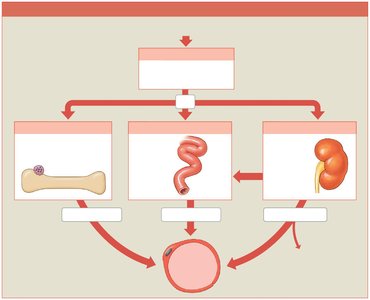
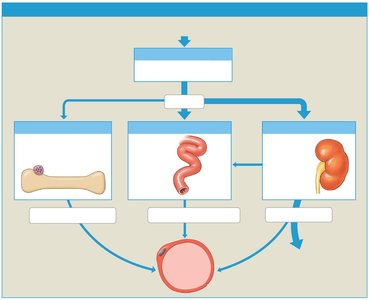
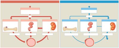
Calcium levels in the blood are tightly regulated by hormonal feedback mechanisms involving the parathyroid and thyroid glands. These mechanisms ensure proper bone health and metabolic function.
Low Blood Calcium: Parathyroid hormone (PTH) is released, stimulating osteoclasts, increasing intestinal absorption, and reducing urinary excretion.
High Blood Calcium: Calcitonin is released, inhibiting osteoclasts, decreasing intestinal absorption, and increasing urinary excretion.
Fractures and Bone Repair
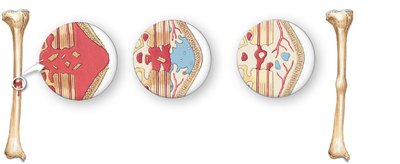
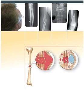
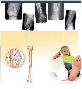
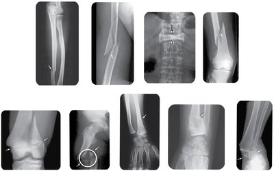
Bone fractures are classified by their characteristics and severity. The repair process involves several stages, including hematoma formation, callus formation, spongy bone formation, and remodeling.
Types of Fractures: Transverse, compression, displaced, nondisplaced, spiral, epiphyseal, comminuted, greenstick, Colles, and Pott's fractures.
Repair Steps: 1. Fracture hematoma formation; 2. Callus formation; 3. Spongy bone formation; 4. Compact bone formation and remodeling.
Bone Disorders: Osteopenia and Osteoporosis
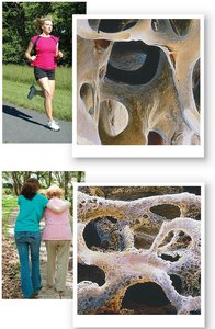
Osteopenia and osteoporosis are conditions characterized by reduced bone mass and increased fracture risk. Osteoclast-activating factor can accelerate bone resorption, contributing to these disorders.
Osteopenia: Mild reduction in bone density, common with aging.
Osteoporosis: Severe reduction in bone density, leading to fragile bones and higher fracture risk.
Osteoclast-activating factor: Increases osteoclast activity, promoting bone resorption.
Additional info:
Vitamin C is necessary for osteoblast differentiation and collagen synthesis.
Vitamins A, K, and B12 are important for bone development and growth in children.
Growth hormone and thyroxine promote protein synthesis and cell metabolism, maintaining the epiphyseal plate.
Testosterone and estrogen accelerate osteoblast activity, resulting in closure of the epiphyseal cartilage and the end of bone growth.
