 Back
Bones and Bone Markings of the Axial and Appendicular Skeleton
Study Guide - Smart Notes
 Tailored notes based on your materials, expanded with key definitions, examples, and context.
Axial Skeleton
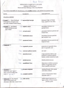
Cranial Bones
The cranial bones form the protective case for the brain and provide attachment points for head and neck muscles. Key cranial bones and their markings include:
Frontal Bone: Forms the forehead and superior part of the orbit.
Supraorbital margin: Marked edge on the frontal bone above the orbit.
Parietal Bone: Dome-shaped bones forming the superior and lateral aspects of the skull.
Occipital Bone: Forms the posterior part of the skull.
Occipital condyles: Articulation between skull and vertebral column.
Foramen magnum: Passage for spinal cord.
Temporal Bone: Located at the sides and base of the skull.
Zygomatic process: Projection to zygomatic bone.
Mastoid process: Attachment for neck muscles.
Styloid process: Attachment for tongue and neck muscles.
Sphenoid Bone: Forms part of the base of the skull.
Sella turcica: Depression housing the pituitary gland.
Ethmoid Bone: Forms part of the nasal cavity and orbital walls.
Facial Bones
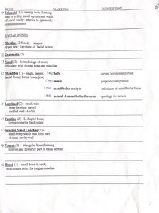
Facial bones shape the face and provide cavities for the sense organs. Important facial bones and their markings include:
Nasal Bone: Forms the bridge of the nose.
Zygomatic Bone: Cheekbone, forms part of the orbit.
Maxilla: Upper jaw, forms part of the hard palate.
Body: Horizontal portion.
Alveolar process: Contains teeth sockets.
Mandibular foramen: Passage for nerves.
Lacrimal Bone: Small bone forming part of the medial wall of the orbit.
Palatine Bone: Forms posterior hard palate.
Inferior Nasal Concha: Forms part of the lateral walls of the nasal cavity.
Vomer: Forms part of the nasal septum.
Hyoid Bone: Located in the neck, supports tongue muscles.
Vertebral Column
Vertebrae
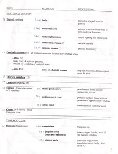
The vertebral column consists of individual vertebrae, each with specific markings for articulation and passage of nerves.
Typical Vertebra:
Body: Thick, disc-shaped anterior portion.
Vertebral arch: Forms posterior part of vertebra.
Vertebral foramen: Central opening for spinal cord.
Transverse process: Extends laterally.
Cervical Vertebrae: All contain transverse foramina for vertebral artery.
Atlas (C1): Lacks body and spinous process; supports the skull.
Axis (C2): Has odontoid process (dens) for rotation.
Thoracic Vertebrae: Articulate with ribs.
Lumbar Vertebrae: Largest vertebrae, support lower back.
Sacrum: Triangular bone formed by fusion of five sacral vertebrae.
Coccyx: Small, triangular bone at the base of the vertebral column.
Thoracic Cage
Sternum and Ribs
The thoracic cage protects vital organs and supports the shoulder girdles and upper limbs.
Sternum: Flat bone forming the anterior midline of the thorax.
Manubrium: Superior portion.
Body: Central portion.
Xiphoid process: Inferior tip.
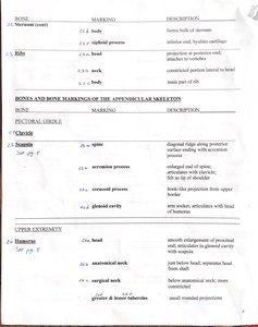
Ribs: Curved bones forming the lateral walls of the thoracic cage.
Head: Articulates with vertebrae.
Neck: Constricted portion between head and body.
Costal cartilage: Connects ribs to sternum.
Appendicular Skeleton
Pectoral Girdle
The pectoral girdle attaches the upper limbs to the trunk and consists of the clavicle and scapula.
Clavicle: S-shaped bone connecting the sternum to the scapula.
Spine: Prominent ridge.
Acromion process: Lateral projection.
Coracoid process: Hook-like projection.
Glenoid cavity: Articulates with humerus.
Upper Extremity
The upper extremity includes the humerus, ulna, radius, carpals, metacarpals, and phalanges.
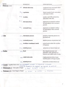
Humerus: Largest bone of the upper limb.
Deltoid tuberosity: Attachment for deltoid muscle.
Capitulum: Articulates with radius.
Trochlea: Articulates with ulna.
Olecranon fossa: Depression for olecranon process.
Ulna: Medial bone of the forearm.
Olecranon process: Forms elbow.
Coronoid process: Anterior projection.
Styloid process: Distal projection.
Radius: Lateral bone of the forearm.
Radial tuberosity: Attachment for biceps brachii.
Styloid process: Distal projection.
Carpals: Eight bones forming the wrist.
Metacarpals: Five bones forming the palm.
Phalanges: Fourteen bones forming the fingers.
Pelvic Girdle
The pelvic girdle attaches the lower limbs to the axial skeleton and supports the weight of the upper body.
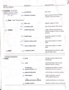
Coxal Bone: Formed by fusion of ilium, ischium, and pubis.
Obturator foramen: Large opening for nerves and blood vessels.
Iliac crest: Superior border of ilium.
Ischial tuberosity: Attachment for hamstring muscles.
Pubic symphysis: Joint between pubic bones.
Lower Extremity
The lower extremity includes the femur, tibia, fibula, tarsals, metatarsals, and phalanges.
Femur: Largest bone in the body.
Head: Articulates with acetabulum.
Neck: Connects head to shaft.
Greater and lesser trochanters: Muscle attachment sites.
Medial and lateral condyles: Articulate with tibia.
Tibia: Medial bone of the leg.
Medial malleolus: Forms inner ankle.
Fibula: Lateral bone of the leg.
Lateral malleolus: Forms outer ankle.
Tarsals: Seven bones forming the ankle.
Metatarsals: Five bones forming the foot.
Phalanges: Fourteen bones forming the toes.
Additional info:
These notes cover the identification and description of major bones and bone markings relevant to the chapters on the skeleton, joints, and the appendicular and axial skeleton. Understanding these structures is essential for anatomy and physiology students, as they form the basis for further study of muscle attachments, joint movements, and clinical applications.
