 Back
BackBones and Bone Structure: Study Notes for ANP College Students
Study Guide - Smart Notes
 Tailored notes based on your materials, expanded with key definitions, examples, and context.
Bones and Bone Structure
Introduction to the Skeletal System
The skeletal system forms the structural framework of the body and is essential for movement, protection, and physiological regulation. It is divided into two main divisions: the axial and appendicular skeletons.
Axial skeleton: Consists of 80 bones including the skull, thorax, and vertebral column. It forms the longitudinal axis of the body.
Appendicular skeleton: Composed of 126 bones of the limbs and girdles that attach them to the axial skeleton.
Associated structures: Cartilages, ligaments, and other connective tissues support and connect bones.
Functions of the Skeletal System
Support: Provides structural support for the entire body.
Mineral and lipid storage: Stores calcium, phosphate, and lipids in yellow bone marrow.
Blood cell production: Occurs in red bone marrow (hematopoiesis).
Protection: Shields vital organs (e.g., skull protects brain, ribs protect thoracic organs).
Leverage: Acts as levers for muscle action, enabling movement.
Classification of Bones and Bone Markings
Bone Shapes
Bones are classified by shape, which relates to their function and location.
Flat bones: Thin, parallel surfaces (e.g., cranial bones, sternum, ribs, scapulae).
Sutural bones: Irregular bones between cranial bones; variable in number and shape.
Long bones: Long and slender (e.g., limbs).
Irregular bones: Complex shapes (e.g., vertebrae, pelvis, facial bones).
Sesamoid bones: Small, flat, develop in tendons (e.g., patella).
Short bones: Small, boxy (e.g., carpals, tarsals).
Bone Markings (Surface Features)
Bone markings are anatomical features on bones that serve as attachment points or passageways for structures such as muscles, tendons, ligaments, blood vessels, and nerves.
Elevations/Projections: Sites for muscle, tendon, and ligament attachment or articulation with other bones.
Depressions/Grooves/Tunnels: Pathways for blood vessels and nerves.
Examples of Bone Markings:
Head: Expanded end of a bone forming part of a joint.
Diaphysis: Shaft of a long bone.
Neck: Narrow connection between head and diaphysis.
Process: Any projection or bump.
Tubercle/Tuberosity: Small, rounded/rough projection.
Trochanter: Large, rough projection (femur).
Condyle: Smooth, rounded articular process.
Facet: Small, flat articular surface.
Crest/Line/Spine: Prominent/low ridge or pointed process.
Ramus: Extension making an angle with the rest of the structure.
Canal/Meatus: Large passageway through bone.
Sinus: Air-filled chamber within bone.
Foramen: Small, rounded passageway for vessels/nerves.
Fissure: Elongated cleft or gap.
Sulcus: Deep, narrow groove.
Fossa: Shallow depression.
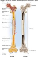
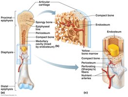
Structure of Long Bones
Gross Anatomy of a Long Bone
Long bones are specialized for transmitting forces and have a complex internal structure.
Epiphysis: Expanded ends, mostly spongy bone, contains red marrow for blood cell production.
Articular cartilage: Covers epiphysis at joints, reduces friction.
Metaphysis: Connects epiphysis to diaphysis.
Diaphysis: Shaft, contains medullary (marrow) cavity with yellow marrow (fat storage).
Periosteum: Outer membrane covering bone except at joints.
Endosteum: Lines medullary cavity and trabeculae of spongy bone.
Microscopic Structure of Bone Tissue
Bone tissue is organized into compact and spongy bone, each with distinct microscopic features.
Osteon (Haversian system): Functional unit of compact bone; concentric lamellae around a central canal.
Lamellae: Layers of bone matrix (concentric, circumferential, interstitial).
Lacunae: Small spaces housing osteocytes.
Canaliculi: Tiny channels connecting lacunae for nutrient/waste exchange.
Perforating canals: Connect central canals of adjacent osteons.
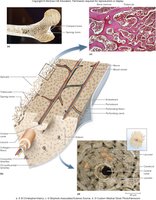
Compact vs. Spongy Bone
Compact bone: Dense, organized into osteons, provides strength for weight bearing.
Spongy bone: Network of trabeculae, lighter, contains red marrow, supports and protects marrow.
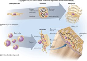
Bone Cells and Bone Matrix
Types of Bone Cells
Osteogenic cells: Stem cells in periosteum and endosteum; differentiate into osteoblasts.
Osteoblasts: Bone-forming cells; secrete bone matrix and become osteocytes.
Osteocytes: Mature bone cells; maintain bone matrix and monitor mineral content.
Osteoclasts: Large, multinucleate cells; break down bone matrix (bone resorption).
Bone Matrix Composition
Collagen fibers: Provide flexibility and tensile strength.
Calcium phosphate: Forms hydroxyapatite crystals, giving bone its hardness and strength.
Other salts: Contribute to bone rigidity.
Bone Growth and Development
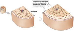
Appositional Growth
Appositional growth increases the diameter of bones by adding new layers of bone matrix under the periosteum.
Osteogenic cells differentiate into osteoblasts, which add circumferential lamellae.
Trapped osteoblasts become osteocytes.
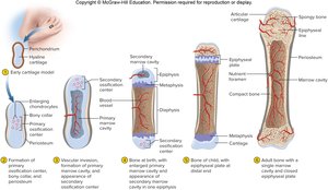
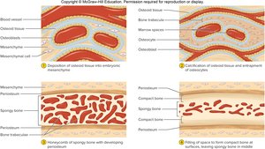
Ossification Processes
Endochondral ossification: Bone replaces hyaline cartilage; forms most bones below the skull except the clavicle.
Intramembranous ossification: Bone develops from fibrous connective tissue; forms flat bones of the skull and clavicle.
Physiology of Bones
Bones as Mineral Reservoirs
Bones store essential minerals, primarily calcium and phosphate, which are vital for physiological processes such as muscle contraction, blood coagulation, and nerve impulse transmission.
Calcium is the most abundant mineral in the body; 99% is stored in the skeleton.
Calcium homeostasis is tightly regulated by the intestines, bones, and kidneys.
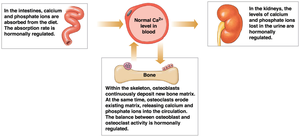
Hormonal Regulation of Calcium
Parathyroid hormone (PTH): Increases blood calcium by stimulating osteoclasts, enhancing intestinal absorption (via calcitriol), and reducing urinary loss.
Calcitonin: Decreases blood calcium by inhibiting osteoclasts, reducing intestinal absorption, and increasing urinary excretion.
Bone Remodeling
Bone remodeling is a continuous process where old bone is replaced by new bone tissue, balancing osteoblast and osteoclast activity.
Bone deposition: Osteoblasts synthesize osteoid, which mineralizes to form new bone.
Bone resorption: Osteoclasts break down bone tissue, releasing minerals into the blood.
Bone Fractures and Repair
Types of Fractures
Closed (simple) fracture: Bone breaks but does not penetrate the skin.
Open (compound) fracture: Bone breaks through the skin; higher risk of infection and bleeding.
Transverse fracture: Break across the long axis.
Spiral fracture: Caused by twisting forces.
Displaced fracture: Bone fragments are misaligned.
Nondisplaced fracture: Bone fragments remain in normal alignment.
Compression fracture: Bone is crushed, often in vertebrae.
Greenstick fracture: Incomplete break, common in children.
Comminuted fracture: Bone shatters into multiple pieces.
Epiphyseal fracture: Occurs at the growth plate; may affect bone growth if not properly managed.
Pott’s fracture: Bimalleolar fracture at the ankle.
Colles fracture: Break in the distal radius.
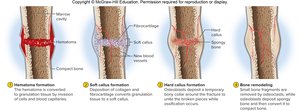
Fracture Healing Process
Bone repair involves four main steps:
Fracture hematoma formation: Blood clot forms at the site, closing injured vessels.
Soft callus formation: Fibrocartilaginous tissue stabilizes the break.
Hard callus formation: Spongy bone replaces the soft callus.
Bone remodeling: Compact bone replaces spongy bone, restoring shape and strength.
Additional info: These notes provide a comprehensive overview of bone structure, function, growth, and repair, suitable for ANP college students preparing for exams or seeking a concise reference.
