 Back
BackBones and Bone Structure: Study Notes for ANP College Students
Study Guide - Smart Notes
 Tailored notes based on your materials, expanded with key definitions, examples, and context.
Chapter 6: Bones and Bone Structure
Introduction to Bone Structure
Bones are dynamic organs that provide support, protection, leverage for movement, storage of minerals, and house bone marrow for blood cell production. Understanding their structure and classification is fundamental in anatomy and physiology.
Classification of Bones by Shape
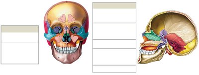
Flat Bones: Thin, parallel surfaces; found in the skull, sternum, ribs, and scapulae. They protect soft tissues and provide surface area for muscle attachment.
Sutural Bones (Wormian Bones): Small, flat, irregular bones between skull bones; variable in number and size.
Irregular Bones: Complex shapes with short, flat, notched, or ridged surfaces; examples include vertebrae and pelvic bones.
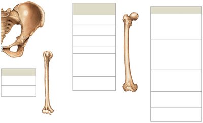
Long Bones: Long and slender; found in limbs (e.g., femur, humerus, phalanges). The femur is the largest and heaviest bone.
Short Bones: Boxlike in appearance; found in wrists (carpals) and ankles (tarsals).
Sesamoid Bones: Small, round, and flat; develop inside tendons near joints (e.g., patella).
Bone Markings
Bone markings are surface features that serve as sites for muscle, tendon, and ligament attachment, or as passages for nerves and blood vessels.
Projections: Sites of muscle and ligament attachment (e.g., trochanter, crest, spine, tubercle, tuberosity).
Projections for forming joints: Head, neck, facet, condyle, trochlea.
Depressions: Sulcus (groove), fossa (shallow depression).
Openings: Foramen (rounded passageway), canal (duct/channel), fissure (slit), meatus (canal-like passage), sinus (air-filled cavity).
Gross Anatomy of Bones
Long Bone Structure
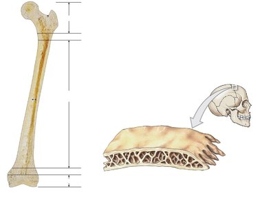
Diaphysis: Shaft; composed of compact bone surrounding the medullary cavity.
Epiphysis: Expanded ends; mostly spongy bone covered by compact bone.
Metaphysis: Region where diaphysis and epiphysis meet; contains the growth plate in children.
Medullary Cavity: Central cavity containing bone marrow.
Flat Bone Structure
Composed of two layers of compact bone with spongy bone (diploë) in between.
Microscopic Structure of Bone
Types of Bone Cells
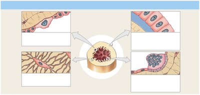
Osteogenic Cells: Stem cells that differentiate into osteoblasts.
Osteoblasts: Immature bone cells that secrete organic components of the matrix (osteoid).
Osteocytes: Mature bone cells that maintain the bone matrix; reside in lacunae and communicate via canaliculi.
Osteoclasts: Large, multinucleate cells that resorb (break down) bone matrix using acids and enzymes.
Matrix Composition: Collagen fibers (provide flexibility and strength) and hydroxyapatite (calcium phosphate + calcium hydroxide; provides hardness).
Compact Bone Structure
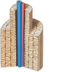
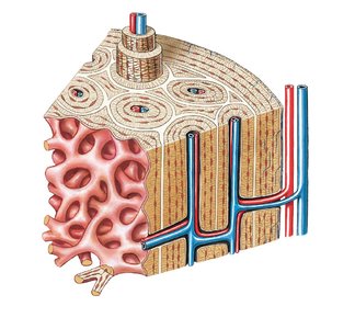
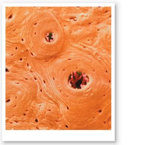
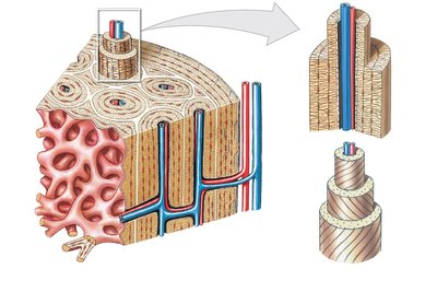
Osteon (Haversian System): The structural unit of compact bone; consists of concentric lamellae around a central canal containing blood vessels and nerves.
Lamellae: Layers of bone matrix.
Lacunae: Small spaces housing osteocytes.
Canaliculi: Tiny channels connecting lacunae for nutrient and waste exchange.
Perforating (Volkmann's) Canals: Transverse canals connecting osteons and medullary cavity to the periosteum.
Spongy Bone Structure
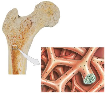
Trabeculae: Network of struts and plates; no osteons present.
Spaces between trabeculae contain red bone marrow (site of hematopoiesis).
Provides strength with minimal weight.
Periosteum and Endosteum
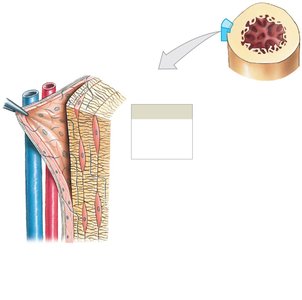
Periosteum: Outer membrane covering bone except at joints; consists of an outer fibrous layer and inner cellular layer. Provides attachment for tendons and ligaments via perforating fibers.
Endosteum: Thin membrane lining the medullary cavity and trabeculae of spongy bone; contains osteogenic cells, osteoblasts, and osteoclasts.
Mechanical Properties of Bone
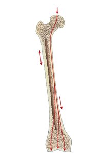
Bones are strong under compression but weaker under tension and shear forces.
Collagen fibers provide flexibility; mineralized matrix provides compressive strength.
Paranasal Sinuses
Air-filled spaces in the skull (frontal, maxillary, sphenoid, ethmoid).
Functions: Lighten the skull, aid in phonation, and can become inflamed (sinusitis).
Summary Table: Types of Bone Cells
Cell Type | Function |
|---|---|
Osteogenic Cell | Stem cell; produces osteoblasts |
Osteoblast | Forms new bone matrix |
Osteocyte | Maintains bone matrix |
Osteoclast | Resorbs bone matrix |
Key Equations
Hydroxyapatite Formation:
Additional info: Bone remodeling is regulated by hormones such as parathyroid hormone (PTH), which stimulates osteoclast activity, and calcitonin, which inhibits osteoclasts and promotes bone formation by osteoblasts.
