 Back
BackBones and Bone Tissue: Structure, Function, and Microscopic Anatomy
Study Guide - Smart Notes
 Tailored notes based on your materials, expanded with key definitions, examples, and context.
Chapter 6: Bones and Bone Tissue
Introduction to Bones as Organs
The skeletal system is composed of bones, cartilage, and joints. Each bone is considered an organ, consisting of osseous tissue, dense regular and irregular connective tissue, and bone marrow. Bones are dynamic, living tissues that play several critical roles in the body.
Functions of the Skeletal System
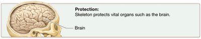
Protection: Bones such as the skull, sternum, ribs, and pelvis protect vital organs from injury.
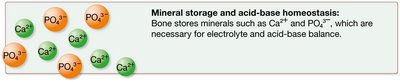
Mineral Storage and Acid–Base Homeostasis: Bones store minerals like calcium, phosphorus, and magnesium, which are essential for electrolyte and acid–base balance.
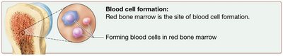
Blood Cell Formation: Red bone marrow is the site of hematopoiesis, the production of blood cells.
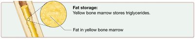
Fat Storage: Yellow bone marrow stores triglycerides, which can be used as an energy source.
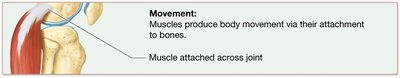
Movement: Bones serve as attachment sites for muscles, enabling movement when muscles contract.
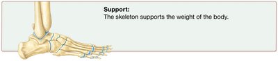
Support: The skeleton provides structural support and bears the weight of the body.
Classification of Bones by Shape
Bones are classified according to their shapes, which relate to their functions:
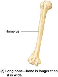
Long Bones: Longer than they are wide; found in arms and legs (e.g., humerus).
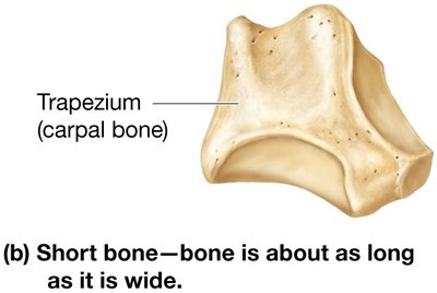
Short Bones: Roughly cube-shaped; found in the wrist (carpals) and ankle (tarsals).
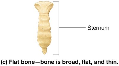
Flat Bones: Thin, broad, and often curved; include ribs, pelvis, sternum, and most skull bones.
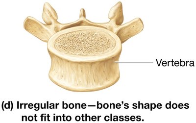
Irregular Bones: Complex shapes that do not fit other categories; include vertebrae and certain skull bones.
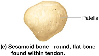
Sesamoid Bones: Small, flat, oval-shaped bones within tendons; provide mechanical advantage (e.g., patella).
Bone Structure
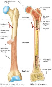
Long Bones: Consist of a diaphysis (shaft), two epiphyses (ends), and a metaphysis (where diaphysis and epiphysis meet). The diaphysis contains the medullary cavity filled with yellow marrow in adults. The epiphyses are covered with articular cartilage and contain spongy bone.
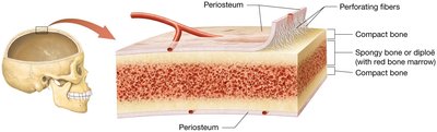
Short, Irregular, and Flat Bones: Composed of thin plates of spongy bone (diploë) covered by compact bone. They lack a diaphysis and epiphyses, and bone marrow is present throughout the spongy bone.
Bone Membranes
Periosteum: Dense irregular connective tissue membrane covering the outer surface of bone; rich in blood vessels and nerves. Perforating (Sharpey's) fibers anchor it to the bone.
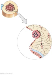
Endosteum: Delicate connective tissue membrane lining internal bone surfaces, covering trabeculae of spongy bone and lining canals in compact bone. Contains osteogenic cells.
Bone Textures
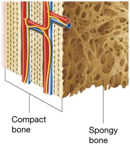
Compact Bone: Dense outer layer that provides strength and resists compression and twisting forces.
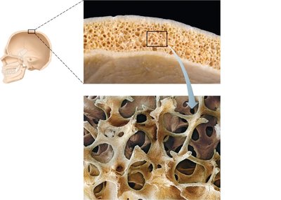
Spongy Bone (Cancellous Bone): Internal honeycomb-like structure that allows resistance to forces from multiple directions and houses bone marrow.
Blood and Nerve Supply
Bones are highly vascularized and innervated. Blood supply to long bones comes from the periosteum and nutrient arteries, which enter through the nutrient foramen. Epiphyses receive blood from small vessels entering through compact bone.
Bone Marrow
Red Bone Marrow: Site of hematopoiesis; found in certain bones in adults (pelvis, proximal femur/humerus, vertebrae, ribs, sternum, clavicles, scapulae, and some skull bones). More abundant in children.
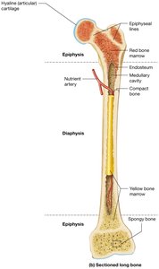
Yellow Bone Marrow: Contains triglycerides, blood vessels, and adipocytes; found in the medullary cavity and some spongy bone spaces in adults.

Microscopic Structure of Bone Tissue
Bone (Osseous) Tissue
Bone tissue is primarily composed of an extracellular matrix with a small population of cells. The matrix is divided into inorganic and organic components.
Extracellular Matrix
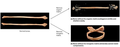
Inorganic Matrix: Makes up about 65% of bone's weight; mainly calcium and phosphorus salts, primarily as hydroxyapatite crystals (). Provides hardness and resistance to compression.
Organic Matrix (Osteoid): Makes up about 35% of bone's weight; consists mainly of collagen fibers, proteoglycans, glycosaminoglycans, glycoproteins, and bone-specific proteins. Provides flexibility and resistance to tensile forces.
Bone Cells
Osteogenic Cells: Stem cells that divide and differentiate into osteoblasts or bone-lining cells.
Osteoblasts: Bone-forming cells that secrete the organic matrix and facilitate mineralization.
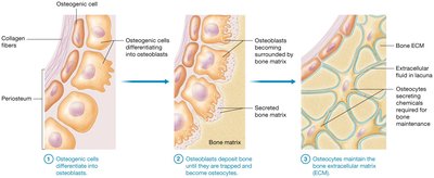
Osteocytes: Mature bone cells that maintain the bone matrix and monitor mechanical stress.
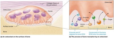
Osteoclasts: Large, multinucleated cells that resorb (break down) bone matrix, derived from hematopoietic stem cells.
Bone-Lining Cells: Flat cells on bone surfaces that help maintain the matrix.
Histology of Compact Bone
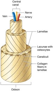
Osteon (Haversian System): The structural unit of compact bone; consists of concentric lamellae (layers of bone matrix) surrounding a central canal containing blood vessels and nerves.
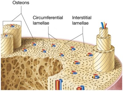
Perforating (Volkmann's) Canals: Run perpendicular to central canals, connecting blood vessels and nerves of periosteum, medullary cavity, and central canal.

Lacunae: Small cavities containing osteocytes.
Canaliculi: Tiny canals connecting lacunae, allowing communication and nutrient/waste exchange between osteocytes.
Interstitial Lamellae: Fill gaps between osteons; remnants of old osteons.
Circumferential Lamellae: Extend around the entire diaphysis, just deep to the periosteum, adding strength and resisting twisting.

Histology of Spongy Bone
Spongy bone is composed of trabeculae (struts or ribs of bone) arranged along lines of stress. It lacks osteons but contains irregularly arranged lamellae and osteocytes interconnected by canaliculi. Capillaries in the endosteum supply nutrients.
*Additional info: Spongy bone lightens the skeleton and provides space for bone marrow, contributing to hematopoiesis and fat storage.*
