 Back
BackBones and Bone Tissue: Structure, Function, and Development
Study Guide - Smart Notes
 Tailored notes based on your materials, expanded with key definitions, examples, and context.
Bones and Bone Tissue
Overview of the Skeletal System
The skeletal system is composed of bones, joints, and supporting tissues. Bones are dynamic organs made up of osseous tissue, connective tissues, and bone marrow. They serve multiple essential functions in the human body.
Functions of the Skeletal System
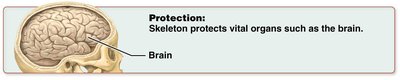
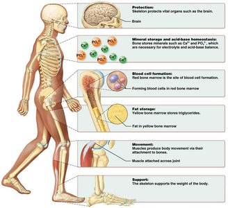
Protection: The skeleton protects vital organs such as the brain, heart, and lungs by encasing them in bone structures (e.g., the skull protects the brain).
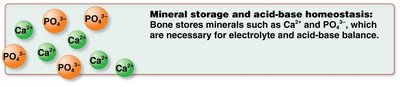
Mineral Storage and Acid-Base Homeostasis: Bones store minerals such as calcium (Ca2+) and phosphate (PO43−), which are essential for electrolyte balance and maintaining acid-base homeostasis in the body.
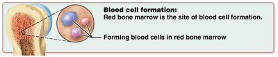
Blood Cell Formation (Hematopoiesis): Red bone marrow is the site of blood cell production, including red blood cells, white blood cells, and platelets.
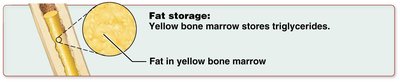
Fat Storage: Yellow bone marrow stores triglycerides, which serve as an energy reserve.
Movement: Bones act as levers for muscles, enabling movement at joints.
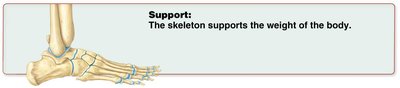
Support: The skeleton provides a structural framework that supports the body’s weight and maintains its shape.
Classification of Bones by Shape
Main Types of Bones
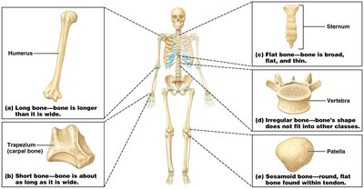
Bones are classified according to their shapes, which relate to their functions and locations in the body.
Long Bones: Longer than they are wide; found in the arms (humerus) and legs (femur).

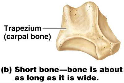
Short Bones: Roughly cube-shaped; found in the wrist (carpals) and ankle (tarsals).
Flat Bones: Thin, broad, and usually curved; found in the skull (parietal), sternum, ribs, and scapulae.
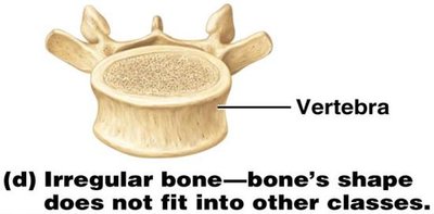
Irregular Bones: Complex shapes that do not fit other categories; found in the vertebrae and certain skull bones.
Sesamoid Bones: Small, round bones embedded within tendons; the patella (kneecap) is the largest example.

Flat Bones (continued): Sternum is an example of a flat bone.
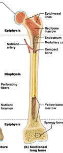
Structure of Long Bones
External and Internal Features
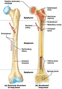
Long bones have a unique structure that supports their function in movement and weight-bearing.
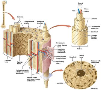
Periosteum: A dense membrane covering the outer surface of bone, containing blood vessels and nerves.
Diaphysis: The shaft or central part of a long bone.
Epiphysis: The expanded ends of the bone, which articulate with other bones.
Articular Cartilage: Hyaline cartilage covering the epiphyses to reduce friction at joints.
Medullary (Marrow) Cavity: Central cavity containing bone marrow (red in children, yellow in adults).
Endosteum: Thin membrane lining the medullary cavity.
Epiphyseal Line/Plate: Remnant of the growth plate, which is a region of hyaline cartilage in growing bones.
Compact and Spongy Bone
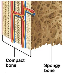
Bone tissue is organized into two main types:
Compact Bone: Dense outer layer that provides strength and resists compression and twisting.
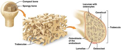
Spongy Bone (Cancellous Bone): Inner, honeycomb-like structure that reduces bone weight and resists forces from multiple directions.
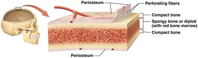
Structure of Other Bone Types
Short, flat, irregular, and sesamoid bones have a simpler structure:
They are covered by periosteum and consist of two thin layers of compact bone with a middle layer of spongy bone (diploë).
Some flat and irregular bones contain sinuses, which are air-filled spaces that reduce bone weight.
Microscopic Structure of Bone Tissue
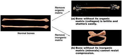
Bone Matrix Composition
The extracellular matrix of bone is composed of:
Inorganic Matrix (65%): Mainly hydroxyapatite crystals (calcium and phosphate), providing hardness and resistance to compression.
Organic Matrix (35%): Called osteoid, consisting of collagen fibers and ground substance, providing flexibility and tensile strength.
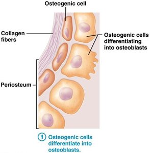
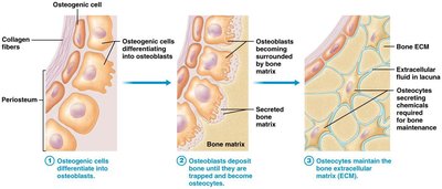
Bone Cells
Osteogenic Cells: Stem cells that differentiate into osteoblasts.
Osteoblasts: Bone-forming cells responsible for bone deposition.
Osteocytes: Mature bone cells that maintain the bone matrix and reside in lacunae.
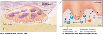
Osteoclasts: Large, multinucleated cells responsible for bone resorption by secreting acids and enzymes.
Histology of Bone
Compact Bone Structure
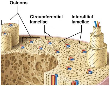
Compact bone is organized into structural units called osteons (Haversian systems):
Lamellae: Concentric rings of bone matrix.
Central Canal: Contains blood vessels and nerves.
Lacunae: Small spaces housing osteocytes.
Canaliculi: Tiny canals connecting lacunae for nutrient and waste exchange.
Perforating (Volkmann’s) Canals: Transverse canals connecting central canals.
Spongy Bone Structure
Spongy bone is composed of a network of trabeculae (bony struts) with spaces containing bone marrow. It is not organized into osteons and is typically found at the ends of long bones and inside flat bones.
Bone Development (Ossification)
Overview of Ossification
Ossification (osteogenesis) is the process of bone formation, beginning in the embryonic period and continuing throughout life. There are two main types of ossification: intramembranous and endochondral.
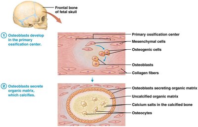
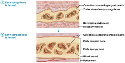
Intramembranous Ossification
This process forms many flat bones, such as those of the skull and clavicles, within a mesenchymal membrane. Spongy bone forms first, followed by compact bone.
Osteoblasts develop in the primary ossification center and secrete organic matrix, which calcifies.
Early spongy bone is formed, followed by early compact bone.
Fontanels are areas of incomplete ossification in the fetal skull.
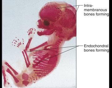
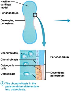
Endochondral Ossification
This process forms most bones of the skeleton (except the clavicles and some skull bones) from a hyaline cartilage model.
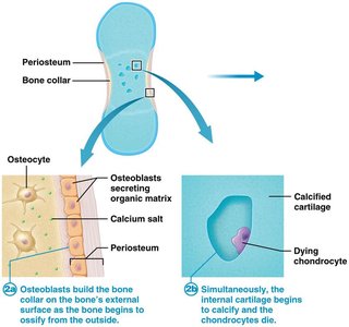
Chondrocytes in the cartilage model die as the matrix calcifies.
Osteoblasts build a bone collar around the diaphysis.
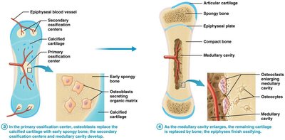
Primary ossification center forms in the diaphysis; secondary centers form in the epiphyses.
Cartilage remains at the epiphyseal plate (growth plate) and articular surfaces.
Summary Table: Bone Types and Examples
Bone Type | Shape/Description | Example |
|---|---|---|
Long | Longer than wide | Humerus, femur |
Short | Cube-shaped | Carpals, tarsals |
Flat | Thin, broad, flat | Sternum, skull bones |
Irregular | Complex shapes | Vertebrae |
Sesamoid | Round, within tendons | Patella |
