 Back
BackBones and Bone Tissue: Structure, Function, and Microscopic Anatomy
Study Guide - Smart Notes
 Tailored notes based on your materials, expanded with key definitions, examples, and context.
Ch. 6 Bones and Bone Tissue
Introduction to Bone and Skeletal Tissue
The skeletal system is composed of bones and associated connective tissues. Bone is a dynamic, living tissue that responds to its environment and plays several critical roles in the body.
Support: Bones provide the structural framework for the body, supporting soft tissues and giving shape to the organism.
Protection: Bones protect vital organs, such as the brain (protected by the skull) and the heart and lungs (protected by the rib cage).
Movement: Bones act as levers for muscles, enabling movement.
Mineral Storage: Bones store minerals, primarily calcium and phosphate, which can be released into the bloodstream as needed.
Fat Storage: Yellow bone marrow stores triglycerides, serving as an energy reserve.
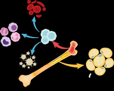
Blood Cell Production: Red bone marrow is the site of hematopoiesis, the production of blood cells.


Types of Bones
Bones are classified based on their shapes and structures, which relate to their functions.
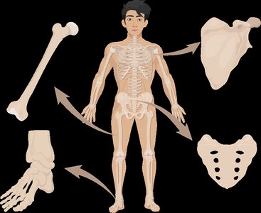
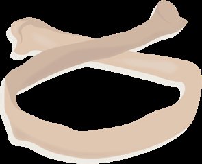
Long Bones: Longer than they are wide, with expanded ends (e.g., femur, humerus). Function as levers for movement.
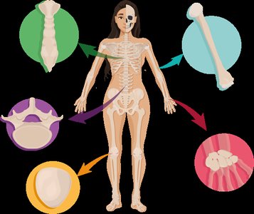
Short Bones: Cube-shaped (e.g., wrist and ankle bones). Provide stability and support with little movement.
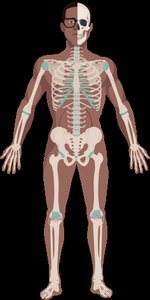
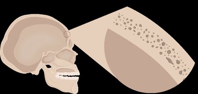
Flat Bones: Thin, flat, and often curved (e.g., sternum, ribs, cranial bones). Provide protection and surfaces for muscle attachment.
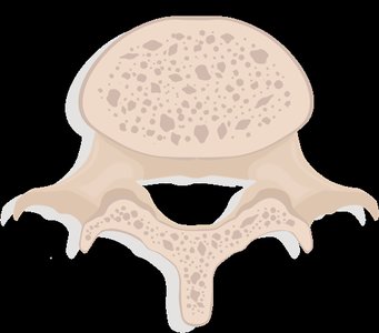
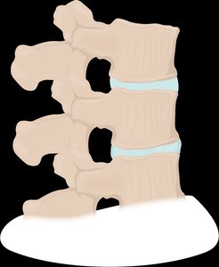
Irregular Bones: Complex shapes (e.g., vertebrae, pelvis, facial bones). Specialized for protection and support.
Sesamoid Bones: Develop within tendons (e.g., patella). Protect tendons from stress and wear.
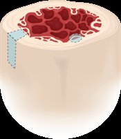
Gross Anatomy of Bone: Compact and Spongy Bone
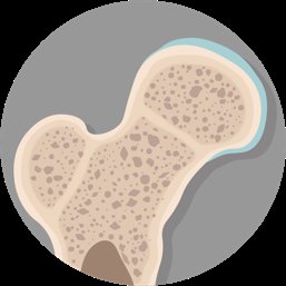
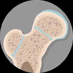
Bone tissue is organized into two main structural types: compact bone and spongy bone.
Compact Bone: Dense and solid, forming the outer layer of all bones and the shaft of long bones. Optimized for strength and hardness, with no visible spaces.
Spongy Bone (Cancellous Bone): Porous, with a lattice-like structure called trabeculae. Found mainly at the ends of long bones and inside flat, short, and irregular bones. Reduces bone weight and contains marrow.
Analogy: Compact bone is like a brick wall (strong and solid), while spongy bone is like scaffolding (lightweight but supportive).


Gross Anatomy of Bone: Periosteum and Endosteum
Bones are covered and lined by specialized connective tissue membranes.
Periosteum: A double-layered membrane covering the external surface of bones (except at joints). The outer fibrous layer is dense irregular connective tissue, while the inner osteogenic layer contains bone stem cells. Perforating fibers anchor the periosteum to the bone matrix, providing strong attachment points for tendons and ligaments.
Endosteum: A thin membrane lining the internal surfaces of bones, including the medullary cavity and trabeculae of spongy bone. Contains osteogenic cells similar to the periosteum's inner layer.

Gross Anatomy of Bone: Bone Marrow
The spaces within bones are filled with bone marrow, which exists in two forms:
Red Marrow: Site of hematopoiesis (formation of blood cells). Found mainly in spongy bone of adults and in most bones of infants and children.
Yellow Marrow: Stores triglycerides (fat). Found in the medullary cavity of long bones in adults. Can convert back to red marrow if necessary (e.g., in cases of severe blood loss).

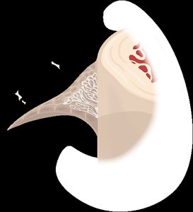

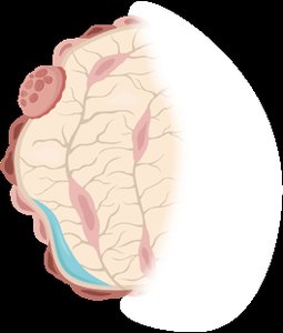
Gross Anatomy of Bone: Structure of Short, Flat, and Irregular Bones
Short, flat, and irregular bones share a similar internal structure: a thin outer layer of compact bone surrounding a core of spongy bone. Flat bones, such as those of the skull, have a "sandwich" structure with compact bone on the outside and spongy bone (diploë) inside.

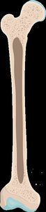
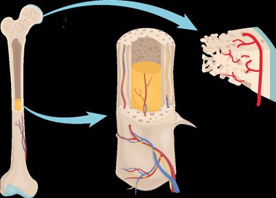
Gross Anatomy of Bone: Structure of a Long Bone
Long bones have a characteristic structure:
Diaphysis: The shaft, composed mainly of compact bone, surrounds the medullary cavity (contains yellow marrow in adults).
Epiphyses: The expanded ends, composed mainly of spongy bone and covered with articular cartilage at joints.
Metaphysis: The region between the diaphysis and epiphysis, containing the epiphyseal plate (growth plate) in children and the epiphyseal line in adults.


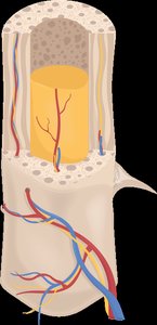
Gross Anatomy of Bone: Nerves and Blood Supply
Bones are highly vascularized and innervated. The nutrient foramen is a small opening in the diaphysis that allows blood vessels and nerves to enter the bone. The nutrient artery supplies the medullary cavity, while veins and nerves exit through the same or additional foramina.
Microscopic Anatomy of Bones: Bone Matrix
The extracellular matrix (ECM) of bone has two main components:
Inorganic Matrix: Composed mainly of hydroxyapatite crystals (calcium and phosphate), making up about 65% of bone mass. Provides hardness and strength for weight-bearing.
Organic Matrix (Osteoid): Composed of collagen fibers and ground substance, making up about 35% of bone mass. Provides flexibility and tensile strength.
Equation for Hydroxyapatite:
Microscopic Anatomy of Bones: Bone Cells
Bone tissue contains several types of cells, each with specialized functions:
Osteoprogenitor (Osteogenic) Cells: Stem cells found in the periosteum and endosteum that differentiate into osteoblasts.
Osteoblasts: Bone-forming cells that secrete the organic matrix (osteoid) and initiate mineralization. Some become trapped in the matrix and differentiate into osteocytes.
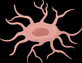
Osteocytes: Mature bone cells that maintain the bone matrix. Located in lacunae and connected by canaliculi for communication and nutrient exchange.
Osteoclasts: Large, multinucleated cells derived from white blood cells. Responsible for bone resorption (osteolysis), breaking down bone matrix using acids and enzymes.


Microscopic Anatomy of Bones: The Osteon (Haversian System)
The osteon is the structural unit of compact bone. Each osteon consists of concentric lamellae (layers of bone matrix) surrounding a central canal that contains blood vessels and nerves. Canaliculi connect lacunae, allowing communication between osteocytes.
Structure | Position | Function | Size |
|---|---|---|---|
Central Canal | Center of osteon; parallel to bone axis | Houses blood vessels and nerves | Large |
Perforating Canal | Perpendicular to central canals | Connects central canal to other vessels/nerves | Large |
Canaliculi | All directions | Communication and transport between osteocytes | Small |
Microscopic Anatomy of Bones: Structure of Lamellae
Lamellae are layers of bone matrix within osteons. Collagen fibers run in alternating directions in adjacent lamellae, providing strength and resistance to twisting forces.
Microscopic Anatomy of Bones: Trabeculae in Spongy Bone
Spongy bone consists of trabeculae—thin, bony struts aligned along lines of stress. Trabeculae contain lamellae, osteocytes, and canaliculi but lack central canals. Nutrients diffuse through canaliculi to reach osteocytes.
Structure | Compact Bone | Spongy Bone |
|---|---|---|
Osteoblasts | Yes | Yes |
Perforating Canals | Yes | No |
Endosteum | Yes | Yes |
Osteons | Yes | No |
Trabeculae | No | Yes |
Osteocytes | Yes | Yes |
Canaliculi | Yes | Yes |
Additional info: The balance between osteoblast and osteoclast activity is essential for bone remodeling and calcium homeostasis. Disorders such as osteoporosis result from an imbalance, leading to weakened bones.
