 Back
BackBones and Organization of the Postcranial Axial Skeleton: Thoracic Cage and Vertebral Column
Study Guide - Smart Notes
 Tailored notes based on your materials, expanded with key definitions, examples, and context.
Bones and Organization of the Postcranial Axial Skeleton
Overview of the Axial Skeleton
The axial skeleton forms the central axis of the human body and includes the skull, vertebral column, and thoracic cage. It provides structural support, protects vital organs, and serves as attachment points for muscles.
Axial Skeleton Components: Skull, vertebral column, thoracic cage (ribs and sternum).
Functions: Protection of the brain, spinal cord, and thoracic organs; support for posture; muscle attachment.
Regions: Cranial, cervical, thoracic, lumbar, sacral, and coccygeal.
Thoracic Cage
The thoracic cage consists of the ribs, sternum, and thoracic vertebrae, forming a protective enclosure for the heart and lungs and facilitating breathing movements.
Structure: Cylindrical, bean-shaped cage with upper limbs dorsal and lower limbs lateral.
Functions:
Protects vital organs (heart, lungs).
Allows movement for breathing via intercostal spaces.
Attachment site for muscles of the neck, back, and shoulders.
Supports pectoral girdle and upper limb attachment.
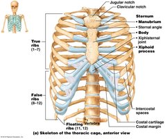
Types of Ribs
There are twelve pairs of ribs, classified based on their attachment to the sternum.
True Ribs (1-7): Attach directly to the sternum via individual costal cartilage.
False Ribs (8-12): Do not attach directly; their costal cartilages merge and attach to the last true rib.
Floating Ribs (11-12): Do not attach to the sternum at all.
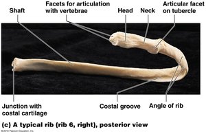
Anatomy of a Typical Rib
Each rib is curved and consists of several anatomical features important for articulation and function.
Shaft: The flat, elongated region.
Costal Groove: Indent on the internal surface for nerves and blood vessels.
Head, Neck, Tubercle: Articulate with thoracic vertebrae.
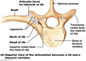
Vertebrocostal Joints
Ribs articulate with thoracic vertebrae at specific points, allowing for movement and stability.
Transverse Costal Facet: Articulates with the tubercle of the rib.
Superior and Inferior Costal Facets: Articulate with the head of the rib.
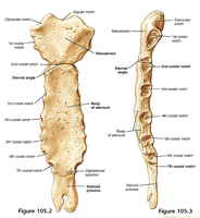
Sternum
The sternum is a flat, T-shaped bone forming the anterior midline of the thoracic cage. It consists of three main parts.
Manubrium: Superior portion; articulates with clavicle and ribs 1-2.
Body: Middle portion; articulates with costal cartilages of ribs 2-7.
Xiphoid Process: Inferior portion; attachment for muscles and costal cartilage.
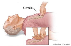
Clinical Implications: CPR
Proper hand placement during cardiopulmonary resuscitation (CPR) is crucial to avoid injury to the xiphoid process and ensure effective compressions.
CPR: Chest compressions at 100-120 per minute keep blood flowing to vital organs.
Hand Placement: Should be over the sternum, not the xiphoid process, to prevent fracture and internal injury.
Vertebral Column
The vertebral column is a series of 24 vertebrae, plus the sacrum and coccyx, providing structural support, protecting the spinal cord, and allowing movement.
Regions: Cervical (7), thoracic (12), lumbar (5), sacrum (5 fused), coccyx (2-6 segments).
Functions: Protects spinal cord, supports posture, muscle attachment, allows flexion, extension, and rotation.
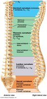
Curvatures of the Vertebral Column
The vertebral column appears straight from anterior/posterior views but has four curvatures in the lateral view.
Primary Curvatures: Thoracic and sacral, present at birth.
Secondary Curvatures: Cervical and lumbar, develop after birth (cervical for head lifting, lumbar for walking).
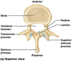
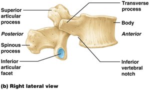
Anatomy of a Typical Vertebra
Each vertebra has several key features that allow for weight-bearing, protection, and movement.
Body: Large, anterior, weight-bearing region.
Vertebral Foramen: Opening for the spinal cord.
Spinous Process: Posterior projection for muscle attachment.
Transverse Processes: Lateral projections for muscle attachment.
Intervertebral Joints and Discs
Intervertebral joints are formed between vertebral bodies and intervertebral discs, which absorb shock and maintain alignment.
Intervertebral Discs: Fibrocartilage pads (23 total) between vertebrae; consist of annulus fibrosus and nucleus pulposus.
Function: Shock absorption, binding vertebrae, allowing limited movement.
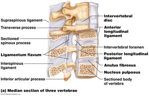
Clinical Implications: Herniated Disc
A herniated (prolapsed) disc occurs when the nucleus pulposus slips out of the annulus fibrosus, pressing on spinal nerves and causing pain.
Symptoms: Pain, numbness, weakness.
Treatments: Exercise, heat therapy, painkillers, or surgical removal.
Comparison of Vertebrae
Vertebrae differ in size, shape, and features depending on their region.
Region | Body Shape | Foramen Shape | Spinous Process | Special Features |
|---|---|---|---|---|
Cervical | Small, oval | Large, triangular | Short, bifid | Transverse foramen |
Thoracic | Heart-shaped | Circular | Long, points inferiorly | Costal facets |
Lumbar | Large, kidney-shaped | Flattened triangle | Thick, flat, posterior | Largest, supports most weight |
Atlas (C1) and Axis (C2)
The first two cervical vertebrae are specialized for head movement.
Atlas (C1): No body or spinous process; superior articular facets for occipital condyles.
Axis (C2): Dens (odontoid process) projects superiorly; allows rotation of the head.
Atlanto-axial Joint: Rotation (“no” movement).
Atlanto-occipital Joint: Flexion/extension (“yes” movement).
Sacrum and Sexual Dimorphism
The sacrum is a fusion of five vertebrae, forming the posterior wall of the pelvis. It exhibits sexual dimorphism.
Male Sacrum: Long and narrow, curves anteriorly.
Female Sacrum: Short and wide, curves posteriorly.
Summary Table: Joints of the Torso
Joint | Bones Involved | Type |
|---|---|---|
Sternoclavicular | Sternum, clavicle | Saddle |
Costosternal | Sternum, ribs (via costal cartilage) | Synchondrosis |
Intervertebral | Vertebral bodies, intervertebral discs | Cartilaginous |
Atlanto-occipital | Atlas, occipital bone | Condyloid |
Atlanto-axial | Atlas, axis | Pivot |
Key Equations and Concepts
Number of Vertebrae:
Number of Ribs:
Additional info:
Embryonic origin of vertebrae and ribs: Derived from the sclerotome portion of somites via endochondral ossification.
Intervertebral discs develop from the notochord and mesenchyme.
