 Back
BackBones and Osseous Tissue: Structure, Function, and Growth
Study Guide - Smart Notes
 Tailored notes based on your materials, expanded with key definitions, examples, and context.
Bones and Osseous Tissue
Functions of Bone
Bones serve several essential functions in the human body, providing structural support, protection, and facilitating movement. They also play a critical role in mineral storage and blood cell production.
Support: Bones form the framework that supports the body and anchors soft tissues.
Protection: Bones protect vital organs, such as the brain (skull), heart and lungs (rib cage), and spinal cord (vertebrae).
Storage of minerals: Bones store minerals, primarily calcium and phosphate, which are released into the bloodstream as needed.
Blood cell production: Red bone marrow within certain bones produces blood cells through hematopoiesis.
Levers for movement: Bones act as levers for muscles, enabling movement.
Bone Tissue: Matrix and Cells
Bone tissue consists of a mineralized matrix and specialized cells that maintain and remodel the bone.
Matrix: Composed of minerals (calcium, phosphates, and other ions) and collagen fibers, providing strength and flexibility.
Bone cells: Responsible for producing, maintaining, and breaking down the matrix during bone remodeling.
Types of Bone/Osseous Tissue
There are two primary types of bone tissue, each with distinct structural and functional characteristics.
Compact bone: Dense and strong, found on the outer parts of bones. The structural unit is the osteon.
Spongy (cancellous) bone: Contains bone marrow and consists of trabeculae, making it lighter and found in areas of low stress.
Types of Bone Cells
Four main types of bone cells contribute to bone formation, maintenance, and remodeling:
Osteocytes: Mature bone cells that maintain the matrix.
Osteoblasts: Cells that build new bone matrix.
Osteoprogenitor cells: Stem cells that differentiate into osteoblasts.
Osteoclasts: Cells that break down bone matrix.
Anatomy of a Long Bone
Structural Features
Long bones have distinct anatomical regions that contribute to their function and growth.
Epiphysis: The ends of the bone, often containing spongy bone and red marrow.
Diaphysis: The shaft of the bone, primarily composed of compact bone.
Metaphysis: The narrow growth zone between epiphysis and diaphysis.
Medullary cavity: The central space in the diaphysis, containing bone marrow.
In adults, the medullary cavity is filled with yellow bone marrow (adipose tissue), while in children it contains red bone marrow (producing blood cells).
Bone Membranes
Bones are covered and lined by specialized membranes:
Periosteum: Covers the outer surface of bone, providing protection and serving as an attachment site for tendons and ligaments.
Endosteum: Lines the inner surface of the medullary cavity, involved in bone growth and remodeling.
Histology of Compact and Spongy Bone
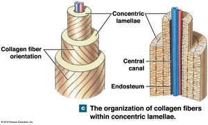
Osteon Structure in Compact Bone
The osteon is the fundamental structural unit of compact bone, consisting of concentric lamellae surrounding a central canal.
Osteocytes: Reside in lacunae and monitor the matrix.
Canaliculi: Tiny channels connecting osteocytes for nutrient and waste exchange.
Central canal: Contains blood vessels and nerves.
Collagen fibers are oriented in different directions in each layer, providing strength and resistance to torsion.
Spongy Bone Structure
Spongy bone consists of a network of trabeculae, making it lighter and suitable for areas of low stress. It contains bone marrow within its spaces.
Bone Development and Ossification
Types of Ossification
Bone formation occurs through two primary processes during embryonic and fetal development:
Intramembranous ossification: Bone develops from a membrane-like layer; forms skull bones.
Endochondral ossification: Bone replaces a cartilage model; forms long bones.
Endochondral Ossification Steps
Endochondral ossification is a multi-step process that transforms cartilage into bone, especially in long bones.
Step 1: Calcification of cartilage matrix kills chondrocytes.
Step 2: Blood vessels grow around cartilage; perichondrium becomes periosteum.
Step 3: Spaces in disintegrating cartilage fill with osteoblasts; spongy bone forms first in diaphysis.
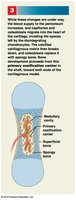
Step 4: Osteoclasts break down matrix to form medullary cavity; osteoblasts form bone in metaphysis for length growth.
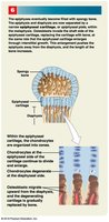
Step 5: Secondary ossification centers form in epiphyses.
Step 6: Cartilage grows at the growth plate (epiphyseal plate); bone is added to diaphysis for lengthening.
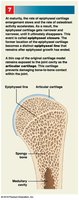
Step 7: Bone stops growing in length at maturity; epiphyseal plate fills with bone, forming the epiphyseal line.
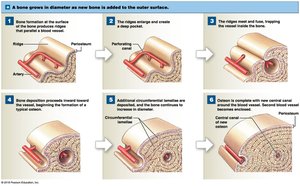
Appositional Growth
Appositional growth refers to the increase in bone diameter as new bone is added to the outer surface.
Osteoblasts add new bone to the periosteum.
Osteoclasts break down bone on the inside, enlarging the medullary cavity.
Regulation of Bone Growth
Minerals and Vitamins
Bone growth and maintenance require a constant supply of minerals and vitamins:
Minerals: Calcium, phosphate, sodium, magnesium, and other ions are essential for matrix hardness.
Vitamin A: Stimulates osteoblast activity.
Vitamin C: Important for collagen synthesis and osteoblast development.
Hormonal Regulation
Several hormones regulate bone growth and remodeling:
Calcitriol and Vitamin D3: Essential for calcium and phosphate absorption in the digestive system. Calcitriol is produced in the kidneys from vitamin D3, which can be synthesized in the skin or obtained from food.
Parathyroid hormone (PTH): Increases blood calcium levels by stimulating osteoclast activity and reducing calcium loss in urine.
Calcitonin: Decreases blood calcium levels by inhibiting osteoclasts; produced in the thyroid gland, especially in children and pregnant women.
Growth hormone and thyroxine: Stimulate bone growth and maintain epiphyseal cartilage activity.
Sex hormones: Estrogen and testosterone accelerate bone growth during puberty, eventually leading to ossification of the epiphyseal plate.
Bone Remodeling and Fracture Repair
Bone Remodeling
Bone remodeling is a continuous process throughout life, balancing osteoblast and osteoclast activity. In young adults, these activities are matched, but in old age, osteoclast activity predominates, leading to bone loss.
Skeleton is regularly replaced.
Bones grow in response to mechanical stress.
Degenerative changes can occur rapidly with age.
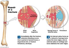
Steps of Fracture Repair
Fracture repair involves several stages to restore bone integrity:
Hematoma formation: A blood clot forms at the fracture site.
Cartilage callus formation: Cartilage stabilizes the fracture.
Bony callus formation: Spongy bone replaces the cartilage callus.
Bone remodeling: Bone is reshaped to its original form.
Additional info: The notes have been expanded with academic context to ensure completeness and clarity for exam preparation.
