 Back
BackBones and Skeletal Tissue: Structure, Classification, and Key Features
Study Guide - Smart Notes
 Tailored notes based on your materials, expanded with key definitions, examples, and context.
Bones and Skeletal Tissue
Bone Structure and Composition
Bones are hard, lightweight composite materials primarily composed of calcium phosphate in the form of calcium hydroxyapatite. They provide structural support, protection, and serve as sites for blood cell formation. Bone tissue can be classified as either compact (cortical) or cancellous (spongy), each with distinct structural and functional properties.
Compact Bone (Cortical): Dense, forms the outer layer of bones, provides strength and protection.
Cancellous Bone (Spongy/Trabecular): Honeycomb-like structure, found at the ends of long bones, contains red marrow for blood cell formation.
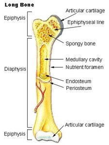
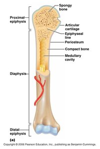
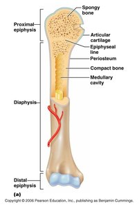
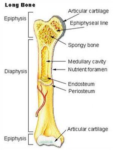
Long Bone Anatomy
Long bones, such as the femur and humerus, are characterized by a shaft (diaphysis) and two ends (epiphyses). They are essential for movement and support.
Epiphysis: The ends of the bone, containing spongy bone and red marrow.
Diaphysis: The shaft, composed mainly of compact bone and housing the medullary cavity with yellow marrow.
Articular Cartilage: Covers joint surfaces, reduces friction and absorbs shock.
Epiphyseal Line: Remnant of the growth plate, important for bone growth during development.
Periosteum: Membrane covering the diaphysis, contains osteoblasts for bone growth and repair.
Endosteum: Membrane lining the medullary cavity.
Histology of Bone: Osteon Structure
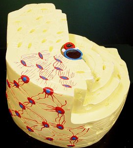
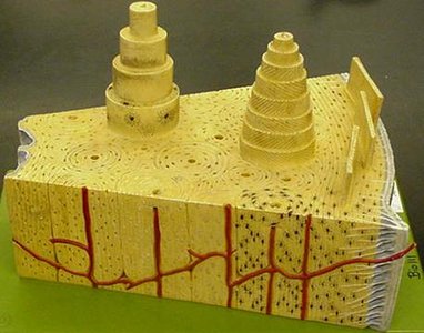
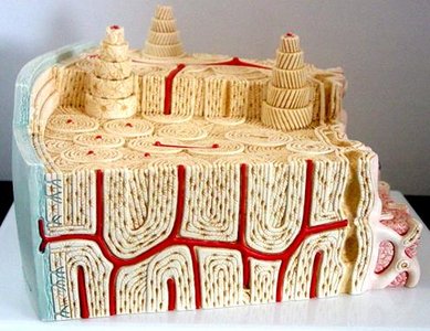
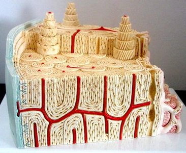
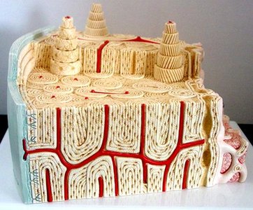
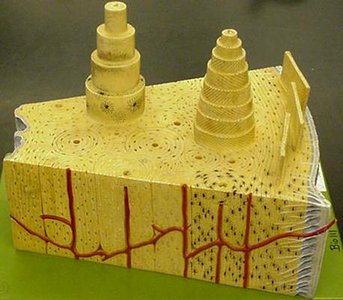
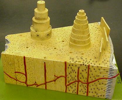
The basic structural unit of compact bone is the osteon (Haversion system). Osteons are cylindrical structures that run parallel to the long axis of the bone, providing strength and facilitating nutrient and waste exchange.
Lamellae: Concentric rings of bone matrix within osteons.
Osteocytes: Mature bone cells located in lacunae (small spaces).
Canaliculi: Tiny channels connecting lacunae, allowing for communication and nutrient/waste exchange between osteocytes.
Central (Haversion) Canal: Vertical canal in the center of each osteon, containing blood vessels, nerves, and lymphatic vessels.
Perforating (Volkmann’s) Canal: Horizontal canals connecting periosteum, central canals, and medullary cavity.
Interstitial Lamellae: Incomplete lamellae between osteons.
Circumferential Lamellae: Lamellae that encircle the entire diaphysis just deep to the periosteum and superficial to the endosteum.
Periosteum and Endosteum
The periosteum is a double-layered membrane covering the external surface of bones (except at joints). It consists of:
Fibrous Layer: Outer layer made of dense connective tissue.
Osteogenic Layer: Inner layer containing osteoblasts (bone-forming cells) and osteoclasts (bone-destroying cells).
Classification of Bones
Bones are classified based on their shape and function:
Long Bones: Shaft and two ends; found in limbs (except ankle, wrist, patella).
Short Bones: Cube-shaped; found in wrist and ankle.
Flat Bones: Thin, flattened, usually curved; found in sternum, scapulae, ribs, skull.
Irregular Bones: Complex shapes; vertebrae, hip bones.
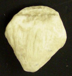
Sesamoid Bones: Formed in tendons; patella.
Bone Surface Markings
Bone surface markings are structural features adapted for specific functions, such as passage of nerves and blood vessels, joint formation, and muscle attachment.
Foramen: Opening or hole.
Fissure: Narrow slit.
Fossa: Shallow depression.
Sulcus: Groove.
Meatus: Tubelike passageway or canal.
Condyle: Large, round protuberance.
Facet: Smooth, flat articular surface.
Trochanter: Very large projection.
Tuberosity: Large, rounded, roughened projection.
Tubercle: Small rounded projection or process.
Fontanels and Sutures of the Skull
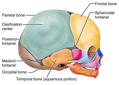
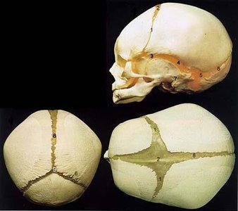
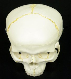
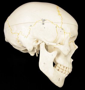
Fontanels are unossified remnants of fibrous membranes in the fetal and infant skull, allowing for brain growth and flexibility during birth. Sutures are immobile joints between skull bones.
Anterior Fontanel: Large, diamond-shaped, closes 6-12 months after birth.
Sphenoidal, Posterior, Mastoid Fontanels: Other soft spots that close early in life.
Sutures: Sagittal, coronal, lambdoid, and squamousal sutures connect various skull bones.
Cranial Bones and Their Features
The skull is composed of several bones, each with unique features and functions:
Frontal Bone: Forms the forehead.
Parietal Bones: Form the superior and lateral aspects of the skull.
Occipital Bone: Forms the posterior wall and base; contains the foramen magnum and occipital condyles.
Temporal Bones: Contain external acoustic meatus, styloid process, mastoid process, zygomatic process, mandibular fossa, carotid canal, jugular foramen.
Zygomatic Bones: Cheekbones; form the zygomatic arch.
Sphenoid Bone: Contains greater and lesser wings, sella turcica, optic canal, foramen rotundum, ovale, spinosum.
Ethmoid Bone: Contains perpendicular plate, conchae, crista galli, cribriform plate.
Maxillae: Upper jaw; contains infraorbital foramen, palatine process.
Lacrimal Bones: House the lacrimal sac.
Nasal Bones: Form the bridge of the nose.
Vomer Bone: Forms part of the nasal septum.
Mandible: Lower jaw; contains coronoid process, mandibular condyle, mental foramen, mandibular foramen, ramus.
Hyoid Bone: Does not articulate with other bones; anchored by ligaments.
The Axial and Appendicular Skeleton
The human skeleton is divided into two main parts:
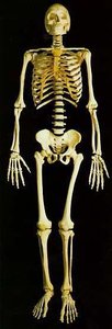
Axial Skeleton: 80 bones; includes skull, hyoid, vertebrae, ribs, sternum, ear ossicles.
Appendicular Skeleton: 126 bones; includes upper and lower limbs, pelvic and pectoral girdles.
Types of Bones: Examples
Long Bone: Femur
Short Bone: Carpal (wrist) bone
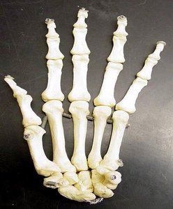
Sesamoid Bone: Patella
Summary Table: Bone Types and Examples
Type | Description | Example |
|---|---|---|
Long | Shaft and two ends | Femur, humerus |
Short | Cube-shaped | Carpals, tarsals |
Flat | Thin, flattened, curved | Sternum, scapula, ribs, skull |
Irregular | Complex shape | Vertebrae, hip bones |
Sesamoid | Formed in tendons | Patella |
Additional info:
Bone histology images reinforce the structure and function of osteons, lamellae, and canals.
Fontanels and sutures are critical for understanding skull development and forensic identification.
Classification and surface markings are essential for anatomical orientation and clinical applications.
