 Back
BackBones and Skeletal Tissues: Structure, Function, and Development
Study Guide - Smart Notes
 Tailored notes based on your materials, expanded with key definitions, examples, and context.
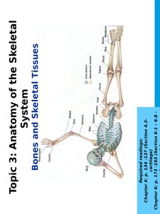
Topic 3: Anatomy of the Skeletal System
Bones and Skeletal Tissues
This topic covers the anatomy and physiology of bones and skeletal tissues, focusing on their structure, classification, growth, and repair. Understanding these concepts is fundamental for students studying human anatomy and physiology.
Learning Objectives
Cartilage
General Features: Cartilage is a tough but flexible connective tissue, with cells (chondrocytes) located in small cavities (lacunae) within a gel-like matrix rich in glycosaminoglycans (GAGs).
Perichondrium: Dense connective tissue layer surrounding cartilage, aids in growth and repair.
Cell Types: Chondroblasts (immature, matrix-secreting cells), Chondrocytes (mature, maintain matrix), Lacunae (spaces housing chondrocytes).
Additional info: Cartilage is avascular, so it heals poorly compared to other tissues.
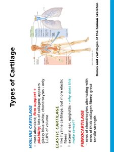
Types of Cartilage
Cartilage is classified based on its fiber content and function:
Hyaline Cartilage: Provides support and flexibility; most abundant type; found in articular surfaces, costal cartilage, nose, trachea.
Elastic Cartilage: More elastic fibers; found in external ear and epiglottis.
Fibrocartilage: Rows of chondrocytes alternating with thick collagen fibers; high tensile strength; found in intervertebral discs and pubic symphysis.
Growth of Cartilage
Appositional Growth: New matrix laid down on surface by perichondrium.
Interstitial Growth: Chondrocytes divide and secrete new matrix within cartilage.
Cartilage growth ends during adolescence.
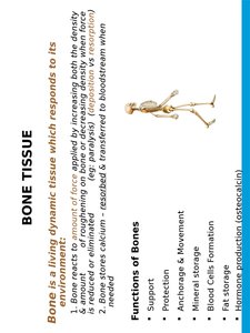
Bone Tissue
Functions of Bones
Bones are living, dynamic tissues that respond to mechanical stress. They serve several essential functions:
Support: Framework for the body.
Protection: Protects vital organs (e.g., skull protects brain).
Anchorage & Movement: Muscles attach to bones for movement.
Mineral Storage: Stores calcium and phosphate.
Blood Cell Formation: Hematopoiesis occurs in bone marrow.
Fat Storage: Yellow marrow stores fat.
Hormone Production: Osteocalcin regulates bone formation and metabolism.
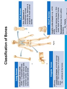
Classification of Bones
Axial vs. Appendicular Skeleton
Axial Skeleton: Skull, vertebral column, rib cage.
Appendicular Skeleton: Limbs and girdles attaching limbs to axial skeleton.
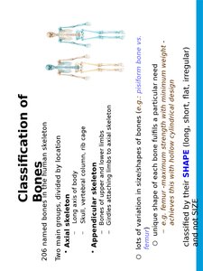
Bone Shapes
Long Bones: Longer than wide; e.g., femur, humerus.
Short Bones: Cube-shaped; e.g., wrist, ankle.
Flat Bones: Thin, flattened; e.g., sternum, scapula.
Irregular Bones: Complex shapes; e.g., vertebrae, hip bones.
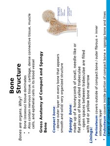
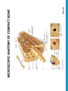
Bone Structure
Gross Anatomy of Compact and Spongy Bone
Compact Bone: Dense outer layer, smooth and solid.
Spongy Bone: Honeycomb of trabeculae (needle-like or flat pieces); spaces filled with bone marrow.
Linings: Periosteum (outer fibrous layer), Endosteum (inner lining).
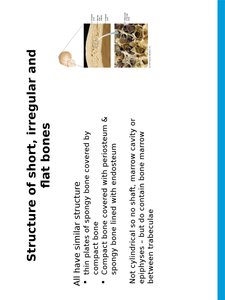
Structure of Short, Irregular, and Flat Bones
Thin plates of spongy bone covered by compact bone.
Covered externally by periosteum, lined internally by endosteum.
No shaft or epiphyses; contain bone marrow between trabeculae.
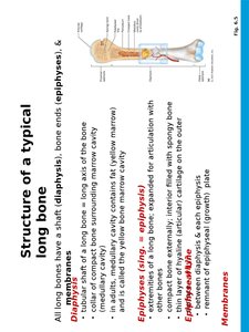
Structure of a Typical Long Bone
Diaphysis: Shaft; tubular, surrounds medullary cavity.
Epiphyses: Bone ends; spongy bone covered by compact bone.
Membranes: Periosteum (outer), endosteum (inner).
Microscopic Anatomy of Bone
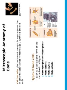
Types of Bone Cells
Osteoprogenitor Cells: Stem cells for bone formation.
Osteoblasts: Bone-forming cells.
Osteocytes: Mature bone cells, maintain matrix.
Osteoclasts: Bone-resorbing cells.
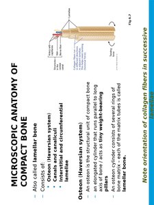
Microscopic Anatomy of Compact Bone
Osteon (Haversian System): Structural unit; consists of concentric lamellae around a central canal.
Canals and Canaliculi: Central canal (blood vessels, nerves), perforating canal (connects osteons), canaliculi (communication between osteocytes).
Lamellae: Collagen fibers in successive layers, resist twisting.
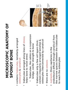
Microscopic Anatomy of Spongy Bone
Contains trabeculae, lamellae arranged along lines of stress.
Osteocytes and canaliculi present; no osteons.
Spaces between trabeculae contain bone marrow.
Chemical Composition of Bone
Organic Components: Cells (osteogenic, osteoblasts, osteocytes, osteoclasts), osteoid (matrix of collagen fibers and ground substance).
Inorganic Components: Hydroxyapatites (mineral salts, mainly calcium phosphate crystals).
Additional info: Bone is half as strong as steel in resisting compression and as strong as steel in resisting tension.
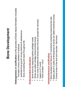
Bone Development
Osteogenesis/Ossification
Formation of bone tissue from embryonic development through childhood and adolescence.
Two main types: Endochondral Ossification (bone forms by replacing hyaline cartilage) and Intramembranous Ossification (bone develops from fibrous membrane).
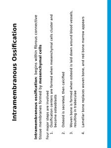
Intramembranous Ossification
Begins within fibrous connective tissue membranes; forms flat bones (e.g., skull, clavicles).
Four steps: mesenchymal cells cluster, osteoid is secreted, woven bone forms, lamellar bone replaces woven bone.
Endochondral Ossification
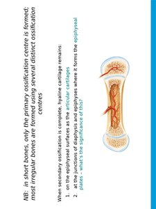
Bone forms by replacing hyaline cartilage; forms most bones below the skull.
Primary ossification center forms in diaphysis; secondary centers in epiphyses.
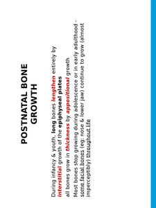
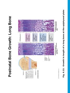
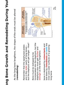
Postnatal Bone Growth
Growth in Length
Long bones lengthen by interstitial growth of epiphyseal plate cartilage.
Epiphyseal plate remains same thickness during childhood; closes at end of adolescence.
Growth ends at about 18 years (female) or 21 years (male).
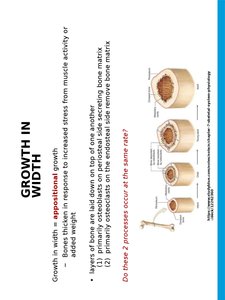
Growth in Width
Bones thicken by appositional growth; layers of bone matrix laid down by osteoblasts.
Osteoclasts remove bone matrix from endosteal surface.
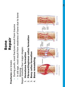
Bone Repair
Fracture Repair
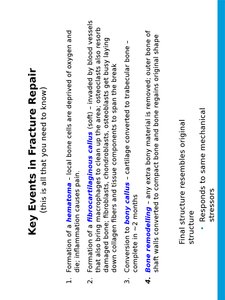
Repair involves four stages: hematoma formation, fibrocartilaginous callus formation, bony callus formation, bone remodeling.
Key Events in Fracture Repair
Hematoma formation: blood clot forms at fracture site.
Fibrocartilaginous callus: cartilage and bone matrix form to stabilize fracture.
Bony callus: new bone replaces callus.
Bone remodeling: bone returns to original shape and structure.
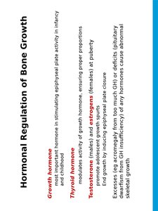
Hormonal Regulation of Bone Growth
Growth Hormone: Stimulates epiphyseal plate activity.
Thyroid Hormone: Modulates activity of growth hormone.
Testosterone and Estrogens: Promote growth spurts and end growth by closing epiphyseal plate.
Bone Modeling and Remodeling
Bone remodeling consists of bone deposit and resorption; occurs throughout life.
Resorption by osteoclasts; deposition by osteoblasts.
About 5% of bone mass is recycled each week.
Control of Remodeling
Regulated by two control loops: maintaining calcium homeostasis (parathyroid hormone and calcitonin) and keeping bone strong (mechanical and gravitational forces).
Additional info: Bone remodeling is essential for bone health, adaptation to stress, and calcium regulation in the body.
