 Back
BackBones and Skeletal Tissues: Structure, Function, and Development
Study Guide - Smart Notes
 Tailored notes based on your materials, expanded with key definitions, examples, and context.
Bones and Skeletal Tissues
Cartilages Help Form the Skeleton
The human skeleton initially consists of cartilage, which is gradually replaced by bone during development. Skeletal cartilage is highly resilient and consists mainly of water, making it flexible yet strong. It lacks blood vessels and nerves, but is surrounded by the perichondrium, a dense connective tissue layer that resists outward expansion and contains blood vessels. Cartilage is composed of chondrocytes housed in lacunae within a jelly-like extracellular matrix.
Structure, Types, and Locations of Cartilage
There are three main types of cartilage in the skeleton:
Hyaline cartilage: Provides support, flexibility, and resilience; contains only collagen fibers.
Elastic cartilage: Contains both collagen and elastic fibers, allowing for greater flexibility.
Fibrocartilage: Contains thick collagen fibers, providing great tensile strength.
Functions of Bones
Major Functions
Bones perform seven essential functions:
Support: Framework for the body.
Protection: Shields vital organs.
Anchorage: Attachment points for muscles.
Mineral storage: Reservoir for calcium and phosphate.
Hematopoiesis: Blood cell formation in red marrow.
Triglyceride storage: Energy storage in yellow marrow.
Hormone production: Osteocalcin regulates insulin secretion, glucose homeostasis, and energy expenditure.
Classification of Bones
Axial and Appendicular Skeleton
The 206 named bones in the human skeleton are divided into two groups:
Axial skeleton: Long axis of the body; includes skull, vertebral column, and rib cage.
Appendicular skeleton: Bones of the limbs and girdles attaching them to the axial skeleton.
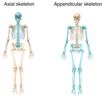
Classification by Shape
Bones are also classified by shape:
Long bones: Longer than wide, with a medullary cavity.
Short bones: Cube-shaped; includes sesamoid bones formed within tendons.
Flat bones: Thin, flat, and slightly curved.
Irregular bones: Complicated shapes not fitting other categories.
Bone Structure
Gross Anatomy of Bone
Bones are organs composed of bone tissue, nervous tissue, cartilage, dense connective tissue, muscle cells, and epithelial cells. There are three levels of structure: gross, microscopic, and chemical.
Compact bone: Dense outer layer, smooth and solid.
Spongy bone: Honeycomb of trabeculae, with spaces filled by red or yellow bone marrow.
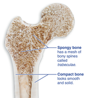
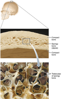
Structure of Short, Irregular, and Flat Bones
These bones consist of thin plates of spongy bone (diploë) covered by compact bone, sandwiched between connective tissue membranes (periosteum and endosteum). Bone marrow is scattered throughout spongy bone, with no defined marrow cavity. Hyaline cartilage is present at movable joints.
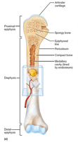
Structure of Typical Long Bone
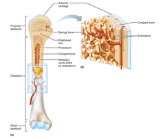
Long bones have a shaft (diaphysis), ends (epiphyses), and membranes:
Diaphysis: Shaft, compact bone surrounding the medullary cavity.
Epiphyses: Ends, compact bone externally and spongy bone internally; articular cartilage covers joint surfaces.
Epiphyseal line: Remnant of the epiphyseal plate, separating diaphysis and epiphysis.
Membranes of Long Bones
Long bones are covered by two membranes:
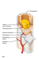
Periosteum: White, double-layered membrane covering external surfaces except joints. The fibrous layer consists of dense irregular connective tissue with Sharpey's fibers, while the osteogenic layer contains stem cells for bone growth.
Endosteum: Covers trabeculae of spongy bone and lines canals in compact bone; contains osteogenic cells.
Hematopoietic Tissue in Bones
Red bone marrow is found within trabeculae of spongy bone and diploë. In newborns, all spongy bone and medullary cavities contain red marrow. In adults, active hematopoiesis occurs mainly in flat bone diploë and some irregular bones (e.g., hip bone). Yellow bone marrow can convert to red marrow under certain conditions, such as increased demand for blood cell production.
Bone Markings
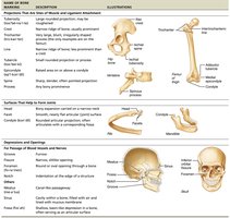
Bone markings are sites for muscle, ligament, and tendon attachment, joint formation, or passageways for blood vessels and nerves. They are categorized as:
Projections: Sites of muscle and ligament attachment.
Surfaces: Form joints.
Depressions and openings: Allow passage of blood vessels and nerves.
Category | Description | Illustration |
|---|---|---|
Projections | Sites for muscle and ligament attachment | Examples: tuberosity, crest, trochanter |
Surfaces | Form joints | Examples: head, facet, condyle |
Depressions and Openings | Passageways for blood vessels and nerves | Examples: foramen, fossa, groove |
Microscopic Anatomy of Bone
Cells of Bone Tissue
Bone tissue contains five major cell types:
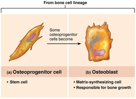
Osteoprogenitor (osteogenic) cells: Stem cells in periosteum and endosteum; differentiate into osteoblasts or bone-lining cells.
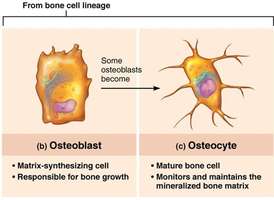
Osteoblasts: Bone-forming cells that secrete unmineralized bone matrix (osteoid); actively mitotic.
Osteocytes: Mature bone cells in lacunae; maintain bone matrix and act as stress sensors.
Bone-lining cells: Flat cells on bone surfaces; help maintain matrix.
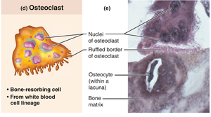
Osteoclasts: Giant, multinucleate cells responsible for bone resorption; derived from hematopoietic stem cells.
Microscopic Anatomy of Compact Bone
Compact bone, also known as lamellar bone, consists of:
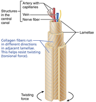
Osteon (Haversian system): Structural unit; elongated cylinder parallel to bone axis; composed of concentric rings (lamellae) with collagen fibers running in different directions to resist twisting.
Canals and canaliculi: Central (Haversian) canal contains blood vessels and nerves; perforating (Volkmann's) canals connect periosteum, medullary cavity, and central canal.
Lacunae: Small cavities containing osteocytes.
Canaliculi: Hairlike canals connecting lacunae, enabling communication and nutrient/waste exchange.
Interstitial and circumferential lamellae: Fill gaps between osteons and extend around the diaphysis, helping resist twisting.
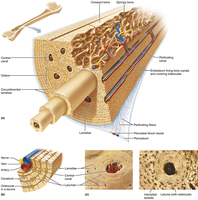
Microscopic Anatomy of Spongy Bone
Spongy bone appears disorganized but is structured along lines of stress. Trabeculae confer strength, and lamellae and osteocytes are interconnected by canaliculi. Capillaries in the endosteum supply nutrients.
Chemical Composition of Bone
Organic Components
Organic components include bone cells and osteoid (unmineralized matrix), which consists of ground substance and collagen fibers. Osteoid provides flexibility and tensile strength.
Inorganic Components
Inorganic components are mainly hydroxyapatites (mineral salts), primarily calcium phosphate crystals. These account for 65% of bone mass and provide hardness and resistance to compression.
Bone Development
Ossification (Osteogenesis)
Ossification is the process of bone tissue formation. The bony skeleton begins forming in the second month of development, and bone growth continues until early adulthood. Bone remodeling and repair are lifelong processes.
Formation of the Bony Skeleton
Up to week 8, the fetal skeleton consists of fibrous membranes and hyaline cartilage, which are replaced by bone tissue via two processes:
Endochondral ossification: Bone forms by replacing hyaline cartilage; forms most of the skeleton.
Intramembranous ossification: Bone develops from fibrous membrane; forms flat bones of the skull and clavicle.
Endochondral Ossification
Forms most bones below the skull (except clavicles). Begins at the primary ossification center in the shaft, where blood vessels infiltrate the perichondrium, converting it to periosteum. Mesenchymal cells become osteoblasts.
Intramembranous Ossification
Begins within fibrous connective tissue membranes formed by mesenchymal cells. Forms frontal, parietal, occipital, temporal, and clavicle bones.
Postnatal Bone Growth
Growth in Length of Long Bones
Long bones grow in length via interstitial growth of the epiphyseal plate, which consists of five zones:
Resting zone
Proliferation (growth) zone
Hypertrophic zone
Calcification zone
Ossification zone
Growth in Width (Thickness)
Bones widen through appositional growth, which can occur throughout life. Osteoblasts secrete bone matrix on the external surface, while osteoclasts remove bone from the internal surface.
Hormonal Regulation of Bone Growth
Key Hormones
Growth hormone: Stimulates epiphyseal plate activity.
Thyroid hormone: Modulates growth hormone activity.
Testosterone and estrogens: Promote adolescent growth spurts and end growth by closing the epiphyseal plate.
Bone Remodeling
Bone Remodeling Process
Bone remodeling replaces 5–10% of bone annually and consists of bone deposit (by osteoblasts) and bone resorption (by osteoclasts). Remodeling occurs at periosteum and endosteum surfaces and is regulated by hormonal and mechanical factors.
Hormonal Control
Blood calcium levels are maintained by:
Parathyroid hormone (PTH): Stimulates osteoclasts to release calcium into the blood.
Calcitonin: Lowers blood calcium levels at high doses.
Mechanical Control
Bone remodeling is also influenced by mechanical stress (Wolff's law): bones grow or remodel in response to the demands placed on them.
Bone Repair
Fracture Classification
Fractures are classified by:
Position of bone ends (nondisplaced vs. displaced)
Completeness of break (complete vs. incomplete)
Whether skin is penetrated (open vs. closed)
Common Types of Fractures
Type | Description |
|---|---|
Comminuted | Bone fragments into three or more pieces |
Compression | Bone is crushed |
Spiral | Ragged break due to twisting forces |
Epiphyseal | Epiphysis separates from diaphysis |
Depressed | Broken bone portion pressed inward |
Greenstick | Bone breaks incompletely |
Fracture Treatment and Repair
Repair involves four stages:
Hematoma forms: Blood vessels hemorrhage, forming a mass of clotted blood.
Fibrocartilaginous callus forms: Capillaries grow into hematoma; fibroblasts secrete collagen fibers; osteoblasts form spongy bone.
Bony callus forms: New trabeculae appear, forming a bony callus.
Bone remodeling: Excess material is removed, and compact bone reconstructs shaft walls.
Bone Disorders
Osteomalacia and Rickets
Osteomalacia: Poorly mineralized bones; soft and weak due to inadequate calcium deposition. Rickets: Osteomalacia in children, resulting in bone deformities; caused by vitamin D deficiency or insufficient dietary calcium.
Osteoporosis
Bone resorption exceeds deposit, leading to decreased bone mass. Most common in aged, postmenopausal females. Treatment includes calcium, vitamin D, exercise, and medications such as bisphosphonates.
Paget’s Disease
Characterized by excessive and disorganized bone remodeling, resulting in weakened bone structure.
Developmental Aspects of Bone
Embryonic and Postnatal Development
The embryonic skeleton ossifies predictably, allowing fetal age determination. Most long bones begin ossifying by 8 weeks, with primary ossification centers developed by week 12. At birth, most long bones are ossified except at epiphyses. Epiphyseal plates persist through childhood and adolescence, and complete ossification occurs by age 25.
