Back
Bones, Joints, and Anatomical Movements: Study Guide for Anatomy & Physiology
Study Guide - Smart Notes
 Tailored notes based on your materials, expanded with key definitions, examples, and context.
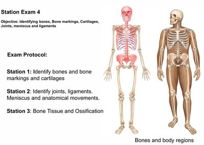
Bones and Body Regions
Overview of the Skeletal System
The human skeletal system provides structure, protection, and support for the body, and serves as the framework for movement. It is composed of bones, cartilage, joints, and associated structures such as ligaments and menisci.
Bones: Rigid organs that form the skeleton and protect internal organs.
Cartilage: Flexible connective tissue found in joints, ear, nose, and other areas.
Joints: Articulations where two or more bones meet, allowing for movement and flexibility.
Ligaments: Strong bands of connective tissue that connect bones and stabilize joints.
Meniscus: Crescent-shaped cartilage structures that improve joint stability and cushioning, especially in the knee.
Bone Markings and Features
Types of Bone Markings
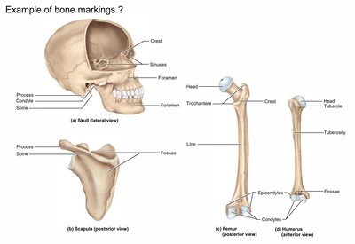
Bones have various markings that serve as attachment points for muscles, passageways for nerves and blood vessels, and articulation surfaces for joints.
Crest: A prominent ridge or elongated projection.
Process: Any bony prominence.
Condyle: A rounded articular projection.
Spine: A sharp, slender projection.
Foramen: A round or oval opening through a bone.
Fossa: A shallow depression or hollow.
Head: The expanded articular end of an epiphysis, separated from the shaft by a neck.
Epicondyle: Raised area on or above a condyle.
Tuberosity/Tubercle: Rounded projection for muscle attachment.
Development of the Skull: Fontanelles
Fontanelles in the Fetal Skull
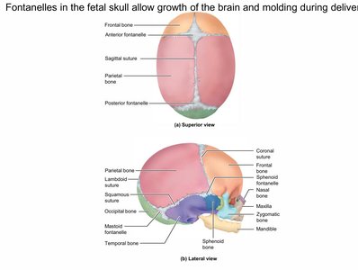
Fontanelles are soft spots on a baby's skull that allow for growth of the brain and facilitate molding during childbirth. These membranous areas eventually ossify as the child grows.
Anterior Fontanelle: Located at the junction of the frontal and parietal bones.
Posterior Fontanelle: Located at the junction of the parietal and occipital bones.
Sphenoid and Mastoid Fontanelles: Located at the sides of the skull.
Vertebral Column and Intervertebral Discs
Structure and Function
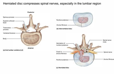
The vertebral column protects the spinal cord and supports the head and body. Intervertebral discs act as shock absorbers and allow flexibility.
Vertebrae: Individual bones of the spine, each with a body, vertebral foramen, and various processes.
Intervertebral Disc: Composed of an outer annulus fibrosus and an inner nucleus pulposus.
Herniated Disc: Occurs when the nucleus pulposus protrudes through the annulus fibrosus, potentially compressing spinal nerves and causing pain, especially in the lumbar region.
Specialized Vertebrae: Atlas and Axis
Atlantoaxial Joint
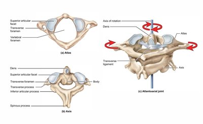
The atlas (C1) and axis (C2) are the first two cervical vertebrae, specialized for supporting the skull and allowing head rotation.
Atlas (C1): Supports the skull; lacks a body and spinous process.
Axis (C2): Has a prominent dens (odontoid process) that acts as a pivot for rotation.
Atlantoaxial Joint: Permits rotation of the head (e.g., shaking head "no").
Regional Differences in Vertebrae
Cervical, Thoracic, and Lumbar Vertebrae
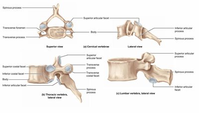
Vertebrae differ in structure depending on their region, reflecting their function and the loads they bear.
Cervical Vertebrae: Small body, large vertebral foramen, transverse foramina.
Thoracic Vertebrae: Larger body, facets for rib articulation, long spinous process.
Lumbar Vertebrae: Largest body, short and thick processes, designed for weight bearing.
Sacrum and Coccyx
Structure and Function
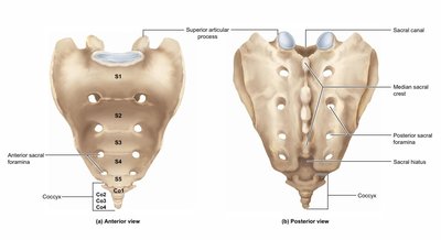
The sacrum is a triangular bone formed by the fusion of five sacral vertebrae, connecting the spine to the pelvis. The coccyx, or tailbone, is composed of fused vertebrae at the base of the spine.
Sacral Canal: Passageway for nerves.
Median Sacral Crest: Fused spinous processes.
Sacral Foramina: Openings for nerves and blood vessels.
Bone Structure: Compact and Spongy Bone
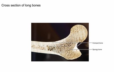
Cross Section of Long Bones
Long bones consist of an outer layer of compact bone and an inner region of spongy bone, which contains red or yellow marrow.
Compact Bone: Dense and strong, forms the outer layer.
Spongy Bone: Porous, found at the ends of long bones and inside flat bones.
Bone Formation: Endochondral Ossification
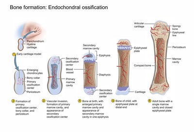
Stages of Endochondral Ossification
Endochondral ossification is the process by which most bones are formed from a cartilage template. This process is essential for the development of long bones.
Formation of cartilage model and perichondrium.
Chondrocytes enlarge, and the matrix calcifies.
Vascular invasion and formation of primary ossification center.
Development of secondary ossification centers in the epiphyses.
Bone growth continues at the epiphyseal plate until adulthood.
Pectoral Girdle: Scapula
Anatomy of the Scapula
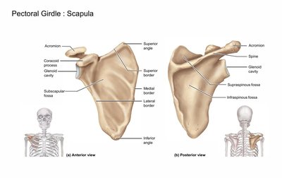
The scapula, or shoulder blade, is a flat bone that forms part of the pectoral girdle. It provides attachment points for muscles and articulates with the humerus and clavicle.
Acromion: Lateral extension of the spine, articulates with the clavicle.
Coracoid Process: Attachment for muscles and ligaments.
Glenoid Cavity: Articulates with the head of the humerus.
Spine: Prominent ridge on the posterior surface.
Joints: Synovial Joints
Structure and Function
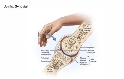
Synovial joints are the most movable type of joint in the body. They are characterized by a joint cavity filled with synovial fluid, articular cartilage, and a fibrous capsule.
Articular Cartilage: Covers bone surfaces, reduces friction.
Synovial Fluid: Lubricates and nourishes the joint.
Ligaments: Strengthen and stabilize the joint.
Anatomical Movements
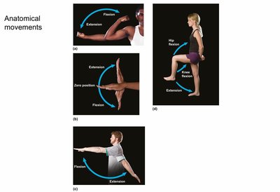
Flexion and Extension
Flexion decreases the angle between two bones, while extension increases it. These movements occur at various joints, including the elbow, knee, and fingers.
Flexion: Bending movement that decreases the angle of the joint.
Extension: Straightening movement that increases the angle of the joint.
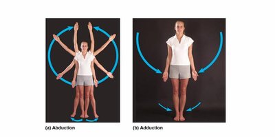
Abduction and Adduction
Abduction is movement away from the midline of the body, while adduction is movement toward the midline.
Abduction: Raising the arms or legs away from the body.
Adduction: Bringing the arms or legs back toward the body.
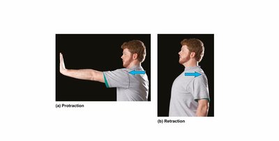
Protraction and Retraction
Protraction moves a body part forward, while retraction moves it backward. These terms are often used for movements of the scapula or jaw.
Protraction: Moving a body part anteriorly (e.g., pushing the jaw forward).
Retraction: Moving a body part posteriorly (e.g., pulling the jaw backward).
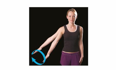
Circumduction and Rotation
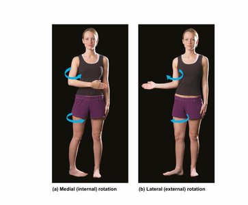
Circumduction is a circular movement that combines flexion, extension, abduction, and adduction. Rotation is movement around a longitudinal axis.
Circumduction: Moving the arm or leg in a circular motion.
Medial (Internal) Rotation: Rotating a limb toward the midline.
Lateral (External) Rotation: Rotating a limb away from the midline.
Supination and Pronation
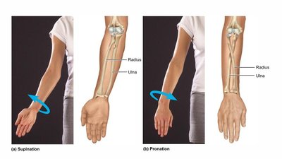
Supination and pronation refer to movements of the forearm. Supination turns the palm upward, while pronation turns it downward.
Supination: Rotating the forearm so the palm faces up.
Pronation: Rotating the forearm so the palm faces down.
Ankle Movements: Dorsiflexion, Plantar Flexion, Inversion, and Eversion
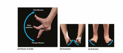
The ankle joint allows for several specific movements:
Dorsiflexion: Lifting the foot upward toward the shin.
Plantar Flexion: Pointing the foot downward.
Inversion: Turning the sole of the foot inward.
Eversion: Turning the sole of the foot outward.
Ligaments and Joint Stability
Role of Ligaments
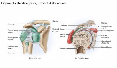
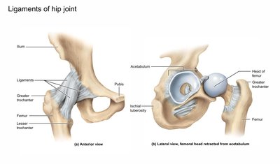
Ligaments are strong, fibrous tissues that connect bones and stabilize joints, preventing dislocations and excessive movement.
Shoulder Joint: Stabilized by ligaments, tendons, and muscles such as the rotator cuff.
Hip Joint: Stabilized by strong ligaments connecting the femur to the pelvis.
Skeletal System: Support and Movement
Functional Importance
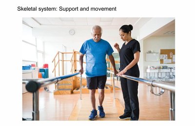
The skeletal system provides the framework for the body, supports movement, and protects vital organs. It works in conjunction with the muscular system to enable locomotion and maintain posture.
Support: Bones provide structural support for the entire body.
Movement: Joints and muscles work together to produce movement.
Protection: Bones protect internal organs such as the brain, heart, and lungs.