 Back
BackBones, Part 1: The Axial Skeleton – Study Notes
Study Guide - Smart Notes
 Tailored notes based on your materials, expanded with key definitions, examples, and context.
The Axial Skeleton
Overview of the Human Skeleton
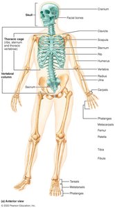
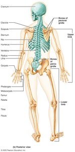
The human skeleton is a structural framework composed of bones, cartilage, joints, and ligaments. It is divided into two main divisions: the axial skeleton and the appendicular skeleton. The axial skeleton consists of 80 bones, including the skull, vertebral column, and thoracic cage, and serves to protect the central nervous system and vital organs.
Axial Skeleton: Skull, vertebral column, thoracic cage
Appendicular Skeleton: Upper and lower limbs, pectoral and pelvic girdles (covered in Chapter 8)
Total Bones: 206 named bones in the adult human body
The Vertebral Column
Structure and Regions
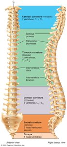
The vertebral column, or spine, is a flexible, curved structure that supports the trunk, protects the spinal cord, and provides attachment points for ribs and muscles. It is divided into five major regions, each with distinct characteristics.
Cervical Vertebrae (C1–C7): Seven vertebrae in the neck
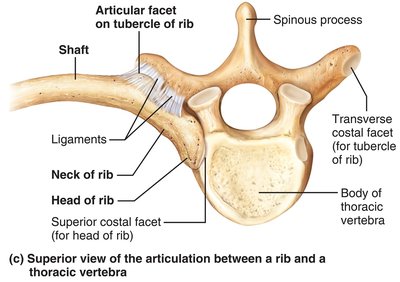
Thoracic Vertebrae (T1–T12): Twelve vertebrae in the upper back, each articulating with ribs
Lumbar Vertebrae (L1–L5): Five vertebrae in the lower back, supporting the most weight
Sacrum: Five fused vertebrae forming the posterior wall of the pelvis
Coccyx: Three to five fused vertebrae forming the tailbone
Normal Curvatures
Cervical and Lumbar Curvatures: Concave posteriorly
Thoracic and Sacral Curvatures: Convex posteriorly
These curvatures increase the spine's resilience and flexibility.
Structure of a Typical Vertebra
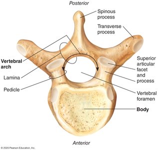
Each vertebra shares a common structural plan, though regional differences exist.
Body: Weight-bearing region
Vertebral Arch: Composed of pedicles and laminae, enclosing the vertebral foramen
Processes: Spinous, transverse, and articular processes for muscle and ligament attachment
Regional Characteristics of Vertebrae
Cervical Vertebrae (C1–C7)
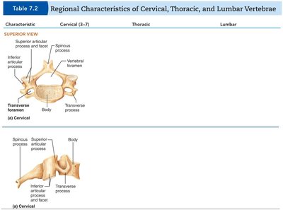
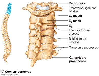
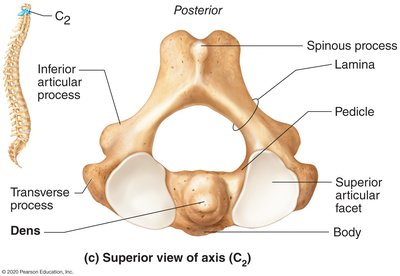
Cervical vertebrae are the smallest and lightest. They have unique features such as transverse foramina and bifid spinous processes (except C7).
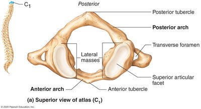
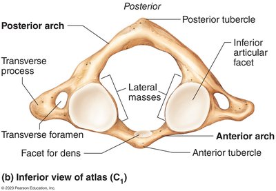
Atlas (C1): Lacks a body and spinous process; supports the skull and allows nodding "yes"
Axis (C2): Has a dens (odontoid process) that acts as a pivot for rotation, allowing the head to indicate "no"
C3–C7: Typical cervical vertebrae with wide bodies and large, triangular vertebral foramina
Thoracic Vertebrae (T1–T12)
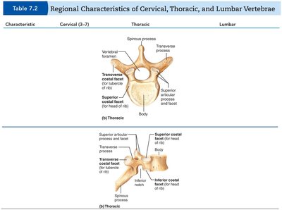
Thoracic vertebrae articulate with ribs and have heart-shaped bodies, long spinous processes, and circular vertebral foramina. Their articular facets allow rotation but limit flexion and extension.
Demifacets: For rib articulation (T1–T10)
Full Facet: T1 for the first rib; T10–T12 have a single facet
Lumbar Vertebrae (L1–L5)
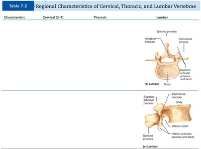
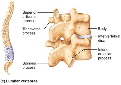
Lumbar vertebrae are large, with thick bodies and short, hatchet-shaped spinous processes. They allow flexion and extension but prevent rotation.
Vertebral Foramina: Triangular
Articular Facets: Oriented medially and laterally
Sacrum and Coccyx
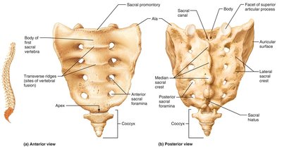
The sacrum forms the posterior wall of the pelvis and is composed of five fused vertebrae. The coccyx, or tailbone, is formed from 3–5 fused vertebrae and provides slight support to pelvic organs.
Sacral Promontory: Anterior border of the first sacral vertebra
Sacral Foramina: Openings for sacral spinal nerves
Ala: Wing-like projections from fused rib elements
The Thoracic Cage
Structure and Function
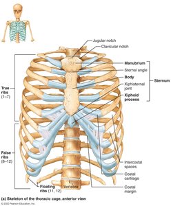
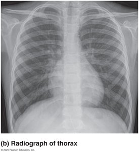
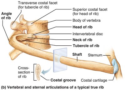
The thoracic cage protects vital organs (heart, lungs) and supports the shoulder girdles and upper limbs. It consists of the sternum, ribs, and thoracic vertebrae.
Sternum: Manubrium, body, and xiphoid process
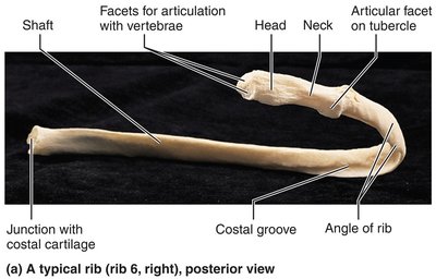
Ribs: 12 pairs, all attaching posteriorly to thoracic vertebrae
True Ribs (1–7): Attach directly to the sternum via costal cartilage
False Ribs (8–12): Attach indirectly or not at all to the sternum; ribs 11–12 are floating ribs
The Skull
Overview and Divisions
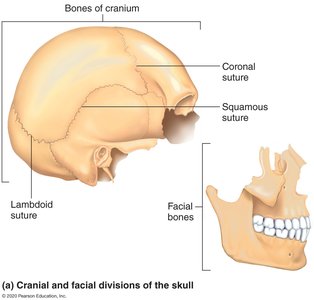
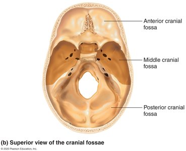
The skull is a complex bony structure that protects the brain and forms the framework of the face. It is divided into cranial and facial bones, with several cavities and numerous foramina for nerves and blood vessels.
Cranial Bones (8): Frontal, parietal (2), temporal (2), occipital, sphenoid, ethmoid
Facial Bones (14): Mandible, vomer, maxillae (2), zygomatic (2), nasal (2), lacrimal (2), palatine (2), inferior nasal conchae (2)
Cavities: Cranial, orbits, nasal, middle and inner ear, paranasal sinuses
Openings: Foramina, canals, and fissures for nerves and vessels
Sutures and Sutural Bones
Sutures are immovable joints connecting the bones of the skull. Four main sutures are present in the cranium, and small, irregular sutural bones may occur within these joints.
Coronal Suture: Parietal and frontal bones
Squamous Suture: Parietal and temporal bones
Sagittal Suture: Right and left parietal bones
Lambdoid Suture: Parietal and occipital bones
Sutural Bones: Small, irregular bones within sutures (not present in all individuals)
Developmental Aspects: Skull of a Newborn
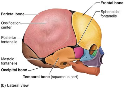
The skull of a newborn features fontanelles—soft spots that allow for brain growth and facilitate passage through the birth canal. These ossify with age.
Anterior Fontanelle: Largest, closes by 18–24 months
Posterior, Mastoid, and Sphenoidal Fontanelles: Close earlier
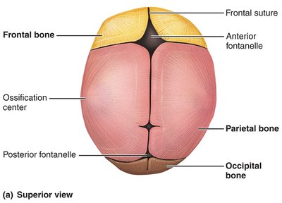
Table: Regional Characteristics of Vertebrae
The following table summarizes the main differences between cervical, thoracic, and lumbar vertebrae:
Characteristic | Cervical (C3–C7) | Thoracic | Lumbar |
|---|---|---|---|
Body | Small, oval, wider side to side | Larger than cervical; heart-shaped | Massive, kidney-shaped |
Vertebral Foramen | Large, triangular | Circular | Triangular |
Spinous Process | Short, bifid (except C7) | Long, sharp, projects inferiorly | Short, blunt, projects directly posteriorly |
Transverse Process | Contain transverse foramina | Bear facets for rib articulation | Thin and tapered |
Articular Facets | Superior face superoposteriorly | Superior face posteriorly | Superior face medially |
Movements Allowed | Flexion, extension, lateral flexion, rotation | Rotation, limited flexion/extension | Flexion, extension; rotation prevented |
