 Back
BackCardiac Contractility and Conduction: Study Notes for ANP College Students
Study Guide - Smart Notes
 Tailored notes based on your materials, expanded with key definitions, examples, and context.
The Heart: Cardiac Contractility and Conduction
Types of Cells in the Heart
The heart contains two main types of cells: autorhythmic fibers and contractile fibers. Each plays a distinct role in cardiac function.
Autorhythmic Fibers: Non-contractile cells (1% of heart cells) that initiate and distribute action potentials (APs). They ensure orderly contraction and set the heart rate. Key components include the SA node, AV node, Bundle of His, bundle branches, and Purkinje fibers.
Contractile Fibers: Cardiac muscle cells (99% of heart cells) that respond to APs and contract to pump blood. These cells are involuntary, short, branched, and contain intercalated discs with gap junctions and desmosomes.
Sequence of Heartbeat: Autorhythmic fibers generate AP → Contractile fibers generate AP → Contractile fibers contract and pump blood.
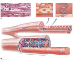
Microscopic anatomy of cardiac muscle showing intercalated discs, gap junctions, and desmosomes.
Microscopic Anatomy of Cardiac Muscle
Cardiac muscle shares similarities with skeletal muscle but has unique features:
Intercalated Discs: Specialized junctions containing gap junctions (allow electrical signals to pass) and desmosomes (provide mechanical strength).
Sarcoplasmic Reticulum: Stores and releases Ca2+.
Mitochondria: Numerous, supporting high energy demand.
T Tubules: Bring excitation to cell interior.
Arrangement of Actin and Myosin: Responsible for contraction.
Action Potentials in Cardiac Muscle
Cardiac muscle cells exhibit a unique action potential sequence, which is longer than that of skeletal muscle.
Resting Membrane Potential (RMP): Negative inside the cell; high K+ inside, high Na+ and Ca2+ outside.
Depolarization: Fast Na+ channels open, Na+ enters cell.
Plateau Phase: Slow Ca2+ channels open, Ca2+ enters cell; some K+ channels open, balancing ion movement.
Repolarization: Ca2+ channels close, K+ channels open, K+ leaves cell.
Extracellular Ca2+ is required for cardiac muscle contraction (unlike skeletal muscle).
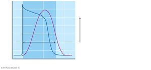
Graph showing the prolonged action potential and contraction in cardiac muscle.
Comparison: Cardiac vs Skeletal Muscle
Cardiac Muscle: AP duration ~200 ms, contraction ~200 ms, refractory period ~200 ms.
Skeletal Muscle: AP duration 1-5 ms, contraction 15-100 ms, refractory period 1-2 ms.
Longer contraction and refractory periods in cardiac muscle ensure effective blood ejection and chamber filling.
Autorhythmic Cells and Pacemaker Function
Autorhythmic cells initiate and distribute impulses throughout the heart, forming the intrinsic conduction system. They require no nervous input to initiate APs and set the heart rate due to their unstable membrane potential.
Na+ slowly leaks into the cell until threshold is reached (-60 mV to -40 mV).
Pacemaker potential: Slow Na+ channels open, K+ channels close.
Depolarization: Fast Ca2+ channels open, Ca2+ enters cell.
Repolarization: Ca2+ channels close, K+ channels open, K+ leaves cell.
Intrinsic Cardiac Conduction System
The conduction system ensures coordinated contraction of the heart chambers.
SA Node: Pacemaker, initiates impulse (~75x/min).
AV Node: Delays impulse (0.1 s), allows atria to contract before ventricles.
AV Bundle (Bundle of His): Only electrical connection between atria and ventricles.
Bundle Branches: Carry impulse toward apex.
Purkinje Fibers: Depolarize contractile cells of ventricles.
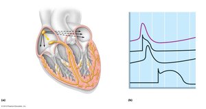
Diagram of the cardiac conduction system showing the sequence of electrical excitation.
Heart Rate Regulation
The fundamental rhythm is set by autorhythmic fibers, but the autonomic nervous system modifies timing and strength.
Parasympathetic (Vagus Nerve): Decreases heart rate.
Sympathetic: Increases heart rate and contractility.
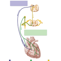
Autonomic innervation of the heart: parasympathetic and sympathetic pathways.
Arrhythmias
Arrhythmias are irregularities in the heart’s rate or rhythm.
Normal Heart Rate: 60-100 beats/min.
Bradycardia: Heart rate below normal range.
Tachycardia: Heart rate above normal range.
Fibrillation: Uncoordinated, rapid contractions; atrial fibrillation (A fib) and ventricular fibrillation (V fib).
Electrocardiography (ECG/EKG)
An ECG records the electrical events of the heart. Key components:
P wave: Atrial depolarization.
QRS complex: Ventricular depolarization (and atrial repolarization).
T wave: Ventricular repolarization.
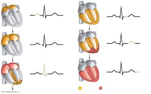
Sequence of depolarization and repolarization of the heart related to ECG tracing.
Cardiac Cycle
The cardiac cycle includes all events associated with blood flow through the heart during one complete heartbeat.
Atrial Systole and Diastole
Ventricular Systole and Diastole
Mechanical events follow electrical events seen in the EKG.
Phases:
Ventricular filling
Isovolumetric contraction
Ventricular ejection
Isovolumetric relaxation
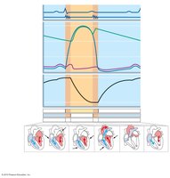
Summary of events during the cardiac cycle: ECG, heart sounds, pressure, and volume changes.
Heart Sounds and Auscultation
Heart sounds are primarily due to valve closure:
1st sound (lub): Closure of AV valves during systole.
2nd sound (dup): Closure of semilunar valves during diastole.
Abnormal sounds (murmurs) may indicate valvular problems.
Cardiac Output
Cardiac output (CO) is the amount of blood pumped by each ventricle per minute.
Formula:
Stroke Volume (SV): Volume of blood pumped by each ventricle per beat.
Formula:
EDV: End Diastolic Volume (amount of blood in ventricle at end of relaxation)
ESV: End Systolic Volume (amount of blood in ventricle at end of contraction)
Factors Affecting Cardiac Output
Heart Rate: Increased HR increases CO.
Stroke Volume: Influenced by preload, contractility, and afterload.
Factor | Definition |
|---|---|
Preload | Degree of stretch on the heart before contraction (determined by EDV) |
Contractility | Force of contraction (increased by Ca2+, sympathetic stimulation, digitalis) |
Afterload | Pressure that must be exceeded before ventricular ejection (increased in hypertension, valvular stenosis) |
Congestive Heart Failure (CHF) and Ejection Fraction
CHF occurs when cardiac output is insufficient for tissue perfusion. Severity is measured by ejection fraction (EF):
Formula:
Normal EF ~60%
Low EF indicates heart failure
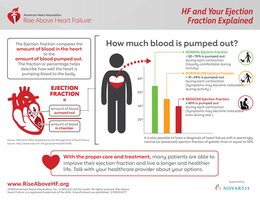
Infographic explaining ejection fraction and its clinical significance in heart failure.
Summary Table: Cardiac Cycle Phases and Valve Status
Phase | Mitral Valve | Aortic Valve | Event |
|---|---|---|---|
Ventricular Filling | Open | Closed | Blood flows from atrium to ventricle |
Isovolumetric Contraction | Closed | Closed | Pressure rises, volume constant |
Ventricular Ejection | Closed | Open | Blood flows from ventricle to aorta |
Isovolumetric Relaxation | Closed | Closed | Pressure falls, volume constant |
Additional info: The notes expand on brief lecture points, providing definitions, formulas, and clinical context for cardiac function and conduction. Images included are directly relevant to the anatomical, physiological, and clinical concepts discussed.
