 Back
BackCardiac Rhythm and Regulation: ANP Study Notes
Study Guide - Smart Notes
 Tailored notes based on your materials, expanded with key definitions, examples, and context.
Cardiac Rhythm and Regulation
Intrinsic Cardiac Conduction System
The heart's rhythm is primarily governed by its intrinsic conduction system, which allows it to contract independently of nervous system input. This system ensures the coordinated contraction of the heart chambers.
Sinoatrial (SA) Node: Known as the pacemaker, located in the right atrial wall. Initiates electrical impulses.
Atrioventricular (AV) Node: Receives signals from the SA node, causing atrial contraction.
Atrioventricular (AV) Bundle: Connects atria to ventricles, transmitting impulses.
Bundle Branches: Conduct impulses through the interventricular septum.
Purkinje Fibers: Depolarize contractile cells in both ventricles, leading to ventricular contraction.
Gap Junctions: Facilitate rapid electrical communication between cardiac cells.
Example: The SA node initiates a heartbeat, which travels through the conduction system, resulting in a coordinated contraction of the heart.
Extrinsic Regulation of Cardiac Rhythm
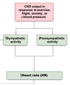
While the heart can beat independently, the autonomic nervous system modulates its rhythm and force of contraction.
Cardioacceleratory Center: Sends signals via the sympathetic trunk to the SA and AV nodes, increasing heart rate and contractility.
Cardioinhibitory Center: Uses the vagus nerve to input directly into the SA and AV nodes, slowing heart rate (parasympathetic effect).
Sympathetic Cardiac Nerves: Increase rate and force of contraction.
Parasympathetic (Vagal Tone): Decreases heart rate by about 25 bpm; cutting the vagal nerve increases HR to ~100 bpm.
Example: During exercise, sympathetic activity increases, raising heart rate and contractility.
Autonomic Nervous System Effects
The autonomic nervous system influences heart rate through neurotransmitters and receptor interactions.
Sympathetic Activation: Triggered by emotional or physical stressors; norepinephrine binds to B1-adrenergic receptors, increasing heart rate and contractility.
Parasympathetic Activation: Acetylcholine opens K+ channels, hyperpolarizing pacemaker cells and slowing heart rate.
Vagal Tone: The heart at rest is under vagal influence, maintaining a lower heart rate.
Example: Anxiety or fright increases sympathetic activity, raising heart rate.
Electrocardiography (ECG)
ECG is a diagnostic tool that records the electrical activity of the heart, providing insight into its rhythm and conduction.
P Wave: Depolarization of the SA node and atria, leading to atrial contraction.
QRS Complex: Ventricular depolarization and atrial repolarization, resulting in ventricular contraction.
T Wave: Ventricular repolarization.
P-R Interval: Time from atrial excitation to ventricular excitation.
S-T Segment: Entire ventricular myocardium depolarized.
Q-T Interval: Duration from ventricular depolarization to repolarization.
Example: Abnormalities in the QRS complex can indicate ventricular conduction issues.
Cardiac Cycle
The cardiac cycle describes the sequence of events during a single heartbeat, including contraction and relaxation phases.
Atrial Systole: Atria contract, pumping blood into ventricles.
Ventricular Systole: Ventricles contract, ejecting blood into circulation.
Diastole: Heart chambers relax, allowing filling with blood.
Example: During diastole, the heart fills with blood, preparing for the next contraction.
Chemical Regulators of Heart Function
Certain hormones and ions play a crucial role in modulating heart rate and contractility.
Epinephrine: Released from the adrenal medulla, increases heart rate and contractility.
Thyroxine: Increases heart rate and enhances the effects of norepinephrine and epinephrine.
Ions: Proper intra- and extracellular concentrations of Ca2+ and K+ are essential for normal heart function.
Example: Hyperkalemia (excess K+) can cause arrhythmias.
Summary Table: Factors Affecting Stroke Volume
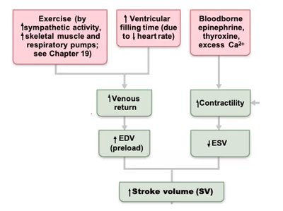
The following table summarizes the main factors influencing stroke volume (SV), which is the amount of blood ejected by the heart per beat.
Factor | Effect on SV | Mechanism |
|---|---|---|
Exercise | Increase | Increases venous return and preload (EDV) |
Sympathetic Activity | Increase | Increases contractility, decreases ESV |
Hormones (Epinephrine, Thyroxine) | Increase | Enhance contractility |
Calcium Ions | Increase | Enhance contractility |
Parasympathetic Activity | Decrease | Reduces heart rate |
Key Equations
Stroke volume and cardiac output are fundamental concepts in cardiac physiology.
Stroke Volume (SV):
Cardiac Output (CO):
Example: If EDV = 120 mL and ESV = 50 mL, then SV = 70 mL.
Additional info: Academic context was added to clarify the conduction system, autonomic regulation, ECG interpretation, and chemical regulation of heart function, as well as to provide relevant equations and a summary table for exam preparation.
