 Back
BackCardiovascular and Lymphatic Systems: Structure and Function
Study Guide - Smart Notes
 Tailored notes based on your materials, expanded with key definitions, examples, and context.
Cardiovascular and Lymphatic Systems
Introduction: The Need for Circulatory Systems
Multicellular organisms require circulatory systems to efficiently transport nutrients, gases, and wastes due to their large size and cell number. Simple diffusion is insufficient for these tasks in larger animals, necessitating specialized transport systems.
Criterion 1: Large number of cells
Criterion 2: Large size and distance from sources of nutrients and oxygen
Key Point: No cell in the human body is more than two cells away from a blood capillary.
Basic Components of Any Circulatory System
All circulatory systems share four fundamental components:
Vehicle: The substance that carries transported materials (e.g., blood or lymph).
Conduits: Tubes or channels through which the vehicle travels (e.g., blood vessels).
Motive Force: The pump or force that moves the vehicle (e.g., heart).
Exchange Areas: Specialized regions where materials are exchanged (e.g., capillaries).
Circulatory Systems in the Human Body
Cardiovascular System: Heart and blood vessels; transports blood.
Lymphatic System: Lymph vessels and nodes; drains interstitial fluid.
Other Minor Systems: E.g., cerebrospinal fluid circulation.
The Human Cardiovascular System
Major Components
The cardiovascular system supplies oxygen and nutrients to cells and removes wastes. Its four major components are:
Blood: The vehicle for transport.
Blood Vessels: The conduits.
Heart: The motive force (pump).
Capillaries: The exchange areas.
Blood: Structure and Function
Blood consists of plasma and formed elements, each with distinct roles.
Plasma: Fluid component (~55% of blood volume), mainly water with dissolved proteins and other substances. After clotting, the remaining fluid is called serum.
Formed Elements: Cellular components (~45% of blood volume):
Red Blood Cells (Erythrocytes): Biconcave discs, contain hemoglobin for oxygen transport. ~5,000,000/mm3.
White Blood Cells (Leukocytes): Immune function. Types include neutrophils (phagocytosis) and lymphocytes (antibody production). ~5,000–11,000/mm3.
Platelets: Cell fragments, aid in clotting. ~150,000–350,000/mm3.
General Functions:
Transport of gases, nutrients, wastes, hormones, antibodies, and heat
Temperature regulation
Immunity (via WBCs and antibodies)
Clotting to prevent blood loss and infection
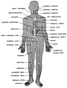
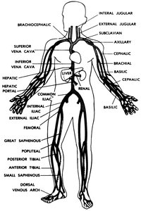
Blood Vessels: Structure and Types
Blood vessels are tubular structures with three-layered walls:
Intima: Inner smooth epithelial lining
Media: Middle layer of smooth muscle
Adventitia: Outer fibrous connective tissue
Three main types of blood vessels:
Arteries: Carry blood away from the heart
Veins: Carry blood toward the heart
Capillaries: Thin-walled vessels for exchange with tissues
Arteries and veins branch into smaller vessels (arterioles and venules) and are largest near the heart. Valves in the heart and veins ensure unidirectional blood flow.
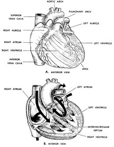
The Heart: Structure and Function
General Construction
The heart is a muscular organ located above the diaphragm, slightly left of center. It is about the size of a clenched fist.
Chambers: Four cavities—right and left atria (upper), right and left ventricles (lower). Auricles are ear-like projections on atria. Septa separate chambers.
Wall Layers:
Endocardium: Inner smooth epithelium
Myocardium: Middle cardiac muscle layer
Epicardium: Outer epithelium
Wall Thickness: Left ventricle wall is thickest, reflecting higher pressure requirements.
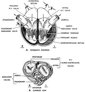
Cardiac Valves
Valves prevent backflow and ensure unidirectional blood movement:
Atrioventricular (A-V) Valves: Between atria and ventricles. Right: tricuspid; left: mitral. Chordae tendineae and papillary muscles prevent valve prolapse.
Semilunar Valves: At bases of major arteries (pulmonary trunk and aortic arch). Pulmonary and aortic semilunar valves have three pocket-like cusps.
Control of the Heart Beat
The heart is regulated by three control systems:
Extrinsic Nervous Control: Autonomic nervous system. Sympathetic nerves accelerate, vagus nerve decelerates heart rate.
Intrinsic "Nervous" Control: Built-in system: sinoatrial (S-A) node (pacemaker), atrioventricular (A-V) node, and septal bundles. Initiates and transmits impulses.
Humoral Control: Blood-borne substances affect heart function, important in transplanted hearts.
Coronary Arteries and Cardiac Veins
The heart receives its own "nutritive" blood supply via the right and left coronary arteries, which arise from the aortic arch. Cardiac veins collect this blood and return it to the right atrium. Blockage of a coronary artery can cause tissue death (myocardial infarction).
Pericardium
The heart is enclosed in the pericardium, a serous sac that reduces friction during contraction.
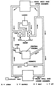
Cardiovascular Circulatory Patterns
General Description
The human cardiovascular system is a closed, two-cycle system:
Closed System: Blood remains within vessels.
Two-Cycle: Blood passes through the heart twice per circuit (pulmonary and systemic cycles).
Collateral Circulation: Multiple vessels supply an area; if one is damaged, others compensate.
End Artery: Single artery supplies a region; damage leads to tissue death.
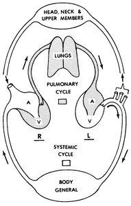
Pulmonary Cycle
Blood flows from the right ventricle through the lungs and returns to the left atrium:
Right ventricle contracts, closing tricuspid valve and opening pulmonary semilunar valve.
Blood enters pulmonary trunk, travels to lungs, exchanges gases in alveolar capillaries.
Oxygenated blood returns via pulmonary veins to left atrium.
Systemic Cycle
Oxygenated blood is pumped from the left ventricle throughout the body and returns deoxygenated to the right atrium:
Left ventricle contracts, closing mitral valve and opening aortic semilunar valve.
Blood enters aortic arch, travels through arteries to tissues.
Exchange occurs in capillary beds; venous system returns blood to right atrium.
Hepatic portal system routes blood from the gut to the liver for processing before returning to circulation.
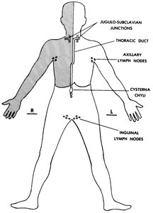
The Human Lymphatic System
General Function
The lymphatic system returns excess interstitial fluid, including proteins, to the bloodstream. It also plays a role in immune defense.
Structures of the Lymphatic System
Lymphatic Capillaries: Absorb excess interstitial fluid in tissue spaces.
Lymph Vessels: Collect lymph; contain valves to ensure one-way flow. The thoracic duct is the major lymph vessel, emptying into the junction of the left subclavian and jugular veins.
Lymph Nodes: Filter lymph fluid; located along lymphatic vessels.
Tonsils: Collections of lymphoid tissue at entrances to respiratory and digestive systems; provide protection.
Summary Table: Components of Circulatory Systems
Component | Cardiovascular System | Lymphatic System |
|---|---|---|
Vehicle | Blood | Lymph |
Conduits | Blood vessels (arteries, veins, capillaries) | Lymphatic vessels |
Motive Force | Heart | Muscle contraction, valves |
Exchange Areas | Capillaries | Lymphatic capillaries, nodes |
Additional info: The notes have been expanded with academic context to clarify the structure and function of each system, and all images included are directly relevant to the adjacent explanations.
