 Back
BackCardiovascular Physiology: Cardiac Muscle, Conduction, and Blood Flow
Study Guide - Smart Notes
 Tailored notes based on your materials, expanded with key definitions, examples, and context.
Cardiovascular Physiology
Overview of the Cardiovascular System
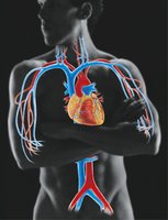
The cardiovascular system is responsible for the circulation of blood throughout the body, delivering oxygen and nutrients to tissues and removing waste products. It consists of the heart, blood vessels, and blood. The heart acts as a pump, propelling blood through a closed network of vessels.
Cardiac Muscle Structure and Function
Microscopic Anatomy of Cardiomyocytes
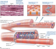
Cardiac muscle cells, or cardiomyocytes, are specialized for continuous rhythmic contraction. They are striated, branched, and typically contain one or two centrally located nuclei. Unique to cardiac muscle are intercalated discs, which contain gap junctions and desmosomes, allowing for electrical and mechanical coupling between cells. This structure enables the myocardium to function as a functional syncytium, meaning the heart contracts as a coordinated unit.
Comparison of Cardiac and Skeletal Muscle
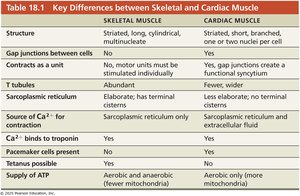
Cardiac and skeletal muscles share some similarities but also have key differences in structure and function. The table below summarizes these differences:
Feature | Skeletal Muscle | Cardiac Muscle |
|---|---|---|
Structure | Striated, long, cylindrical, multinucleate | Striated, short, branched, one or two nuclei per cell |
Gap junctions | No | Yes |
Contracts as a unit | No | Yes |
T tubules | Abundant | Fewer, wider |
Sarcoplasmic reticulum | Elaborate; has terminal cisterns | Less elaborate; no terminal cisterns |
Source of Ca2+ | Sarcoplasmic reticulum only | Sarcoplasmic reticulum and extracellular fluid |
Pacemaker cells | No | Yes |
Tetanus possible | Yes | No |
ATP supply | Aerobic and anaerobic | Aerobic only (more mitochondria) |
Types of Cardiomyocytes
Contractile cells: Make up the majority of the myocardium and are responsible for the heart's pumping action. They have a stable resting membrane potential and require external excitation to contract.
Autorhythmic (pacemaker) cells: Comprise about 1% of cardiac cells and are found in the SA node, AV node, bundle of His, bundle branches, and Purkinje fibers. These cells are self-excitable, have an unstable resting membrane potential, and initiate and propagate action potentials throughout the heart.
Cardiac Conduction System
Intrinsic Electrical Conduction
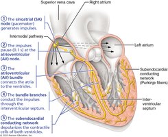
The heart's intrinsic conduction system ensures coordinated contraction. The sequence is as follows:
Sinoatrial (SA) node (pacemaker)
Atrioventricular (AV) node
Atrioventricular (AV) bundle (bundle of His)
Right and left bundle branches
Subendocardial conducting network (Purkinje fibers)
Action Potentials in Cardiac Cells
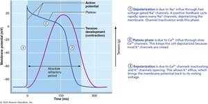
Contractile cells: Exhibit a prolonged action potential with distinct phases: rapid depolarization (Na+ influx), initial repolarization, plateau (Ca2+ influx), and rapid repolarization (K+ efflux).
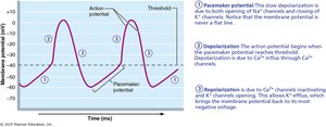
Autorhythmic cells: Display pacemaker potentials due to slow Na+ influx, followed by Ca2+ influx for depolarization, and K+ efflux for repolarization.
Neural Regulation of Heart Rate
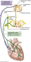
The autonomic nervous system modulates heart rate via the cardiac centers in the medulla oblongata. The parasympathetic system (via the vagus nerve) predominates at rest, slowing the heart rate, while sympathetic stimulation increases heart rate and contractility during stress or exercise.
Electrocardiography (ECG/EKG)
Principles of ECG
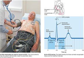
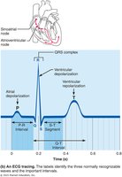
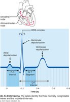
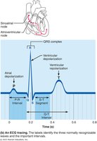
An electrocardiogram (ECG) records the electrical activity of the heart. It consists of characteristic waveforms (P wave, QRS complex, T wave) that correspond to specific electrical events in the cardiac cycle.
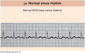
ECG Interpretation and Arrhythmias
Normal sinus rhythm: Regular P, QRS, and T waves.
Junctional rhythm: SA node nonfunctional; AV node paces the heart (absent P waves).
Second-degree heart block: Some SA node impulses not conducted; more P waves than QRS complexes.
Ventricular fibrillation: Disorganized electrical activity; no effective pumping.
Mechanical Events of the Cardiac Cycle
Phases of the Cardiac Cycle
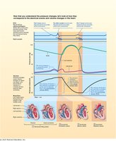
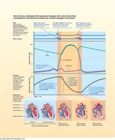
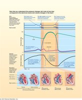
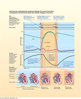
The cardiac cycle consists of alternating periods of contraction (systole) and relaxation (diastole) in the atria and ventricles. Key events include ventricular filling, isovolumetric contraction, ventricular ejection, and isovolumetric relaxation. The Wiggers diagram integrates electrical, pressure, and volume changes during the cycle.
Key Cardiac Volumes and Calculations
End-diastolic volume (EDV): Volume of blood in the ventricle at the end of diastole.
End-systolic volume (ESV): Volume of blood remaining after systole.
Stroke volume (SV): Amount of blood ejected per beat:
Ejection fraction (EF): Percentage of EDV ejected per beat:
Cardiac output (CO): Volume of blood pumped per minute:
Regulation of Cardiac Output
Determinants of Stroke Volume
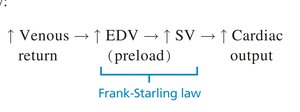
Preload: Degree of stretch of cardiac muscle before contraction (related to EDV). Increased preload increases SV (Frank-Starling law).
Afterload: Pressure the heart must overcome to eject blood (mainly aortic pressure).
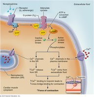
Contractility: Force of contraction at a given preload, influenced by inotropic agents (e.g., Ca2+, norepinephrine).
Heart Rate Regulation
Heart rate is modulated by autonomic innervation, hormones, ions, and other factors. Positive chronotropic agents increase HR, while negative agents decrease it.
Blood Vessels and Circulation
Types and Structure of Blood Vessels
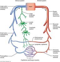
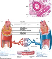
Blood vessels form a closed circuit for blood flow. Major types include arteries, arterioles, capillaries, venules, and veins. Arteries carry blood away from the heart, veins return blood to the heart, and capillaries allow exchange with tissues.
Blood Flow, Pressure, and Resistance
Blood flow (F): Volume of blood moving through a vessel per unit time (ml/min).
Blood pressure (BP): Force per unit area exerted by blood on vessel walls (mmHg).
Resistance (R): Opposition to flow, mainly determined by vessel diameter, length, and blood viscosity.
The relationship is described by:
Where is the pressure gradient and is resistance.
Blood Pressure Regulation
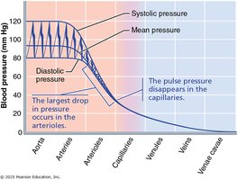
Systolic pressure: Peak pressure during ventricular contraction.
Diastolic pressure: Lowest pressure during ventricular relaxation.
Pulse pressure: Difference between systolic and diastolic pressures.
Mean arterial pressure (MAP): Average pressure driving blood to tissues.
Circle of Willis
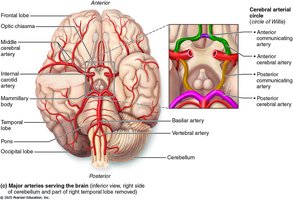
The Circle of Willis is a circular arterial structure at the base of the brain that provides collateral circulation, ensuring constant blood supply to the brain even if one pathway is blocked.
Homeostatic Regulation of Blood Pressure and Flow
Short-Term and Long-Term Regulation
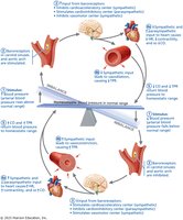
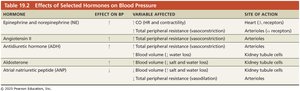
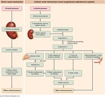
Short-term: Neural (baroreceptor reflex), hormonal (e.g., epinephrine, angiotensin II).
Long-term: Renal mechanisms (regulation of blood volume).
Vasoconstriction and Vasodilation
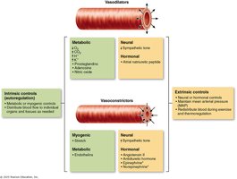
Vasoconstriction increases resistance and blood pressure, while vasodilation decreases resistance and blood pressure. Local and systemic factors regulate vessel diameter to match tissue perfusion needs.
Blood Flow in Capillaries
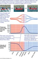
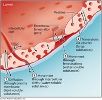
Blood flow is slowest in capillaries due to their large total cross-sectional area, allowing efficient exchange of gases, nutrients, and wastes with tissues.
Clinical Pathologies
Common Cardiovascular Disorders
Hypertension: Chronic high blood pressure, risk factor for heart disease and stroke.
Hypotension: Chronic low blood pressure, may cause dizziness or fainting.
Heart failure: Reduced ejection fraction and cardiac output.
Arrhythmias: Abnormal heart rhythms, including tachycardia, bradycardia, heart block, and fibrillation.
Edema and Capillary Exchange
Edema is the accumulation of excess fluid in tissues, often due to disruption of normal capillary fluid exchange (bulk flow). This can result from increased hydrostatic pressure, decreased oncotic pressure, or lymphatic obstruction.
