 Back
Cardiovascular Physiology: Structure and Function of the Heart
Study Guide - Smart Notes
 Tailored notes based on your materials, expanded with key definitions, examples, and context.
The Heart: Structure and Function
Overview of the Cardiovascular System
The cardiovascular system is responsible for the transport of blood, nutrients, gases, and waste products throughout the body. The heart acts as a central pump, circulating blood through a network of vessels.
Heart Chambers: The heart has four chambers: two atria (upper chambers) and two ventricles (lower chambers).
Blood Flow: Blood flows in a specific sequence through the heart and major vessels, ensuring oxygenation and removal of waste.
Major Vessels: The aorta and pulmonary trunk carry blood away from the heart, while the vena cava and pulmonary veins return blood to the heart.
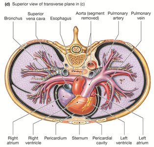
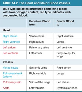
Heart Chambers and Major Blood Vessels
The heart's chambers and associated vessels are organized to maintain efficient circulation. Oxygenated and deoxygenated blood are kept separate to optimize gas exchange.
Right Atrium: Receives deoxygenated blood from the venae cavae.
Right Ventricle: Pumps deoxygenated blood to the lungs via the pulmonary trunk.
Left Atrium: Receives oxygenated blood from the pulmonary veins.
Left Ventricle: Pumps oxygenated blood to the body via the aorta.
Structure | Receives Blood From | Sends Blood To |
|---|---|---|
Right atrium | Venae cavae | Right ventricle |
Right ventricle | Right atrium | Lungs |
Left atrium | Pulmonary veins | Left ventricle |
Left ventricle | Left atrium | Body except for lungs |
Venae cavae | Systemic veins | Right atrium |
Pulmonary trunk (artery) | Right ventricle | Lungs |
Pulmonary vein | Veins of the lungs | Left atrium |
Aorta | Left ventricle | Systemic arteries |
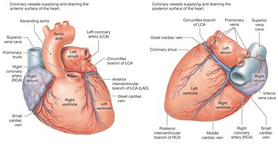
Coronary Circulation
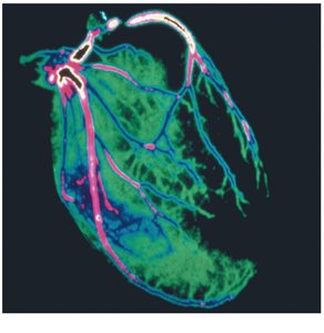
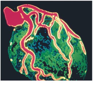
The coronary circulation supplies the myocardium (heart muscle) with oxygenated blood and removes deoxygenated blood. This network is essential for maintaining cardiac function, especially during increased workload.
Coronary Arteries: Deliver oxygen-rich blood to the heart muscle.
Coronary Veins: Remove deoxygenated blood from the myocardium.
Clinical Relevance: Impairment of coronary circulation can lead to myocardial ischemia, arrhythmias, or heart failure.
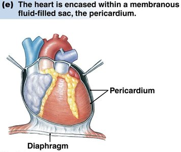
The Pericardium
The pericardium is a strong, membranous sac that encases and protects the heart. It is fused to the diaphragm and contains pericardial fluid, which lubricates the heart and allows it to operate in a friction-free environment.
Function: Reduces friction, protects the heart, and participates in paracrine signaling.
Structure: Composed of fibrous and serous layers.
Ventricular Wall Thickness
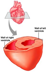
The left and right ventricles have different wall thicknesses due to their distinct functions. The left ventricle has a thicker wall to generate higher pressure for systemic circulation, while the right ventricle pumps blood to the lungs at lower pressure.
Left Ventricle: Thick wall for high-pressure systemic circulation.
Right Ventricle: Thinner wall for low-pressure pulmonary circulation.
Training and the Heart
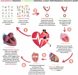
Physical training induces structural adaptations in the heart. Endurance training increases the internal dimensions of the left ventricle, enhancing stroke volume, while resistance training increases wall thickness, improving the heart's ability to pump against higher pressures.
Endurance Training: Enlarges left ventricular cavity, increases stroke volume.
Resistance Training: Increases left ventricular wall thickness, strengthens heart muscle.
Health Impact: Both types of training improve cardiovascular efficiency and reduce disease risk.
Heart Valves and Blood Flow
Heart Valves: Structure and Function
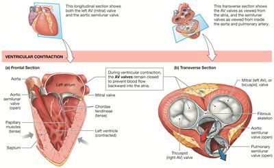
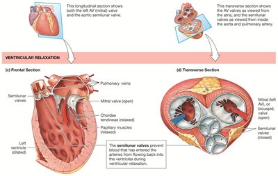
Heart valves ensure one-way flow of blood through the heart. There are two main types: atrioventricular (AV) valves and semilunar valves.
Atrioventricular Valves: Located between atria and ventricles; include the tricuspid (right) and bicuspid/mitral (left) valves.
Semilunar Valves: Located between ventricles and arteries; include the aortic and pulmonary valves.
Chordae Tendineae: Prevent AV valve eversion during ventricular contraction.
Valve Function During Cardiac Cycle
Valves open and close in response to pressure changes during the cardiac cycle, preventing backflow and ensuring efficient circulation.
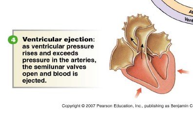
Ventricular Contraction: AV valves close, semilunar valves open.
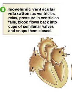
Ventricular Relaxation: Semilunar valves close, AV valves open.
The Cardiac Cycle
Phases of the Cardiac Cycle
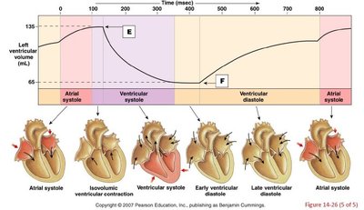
The cardiac cycle consists of a series of events that occur during one heartbeat, including systole (contraction) and diastole (relaxation) of the atria and ventricles.
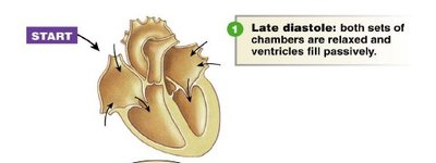
Late Diastole: Both sets of chambers are relaxed; ventricles fill passively.
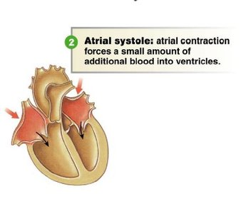
Atrial Systole: Atrial contraction forces additional blood into ventricles.
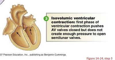
Isovolumic Ventricular Contraction: AV valves close; pressure increases but semilunar valves remain closed.
Ventricular Ejection: Semilunar valves open; blood is ejected into arteries.
Isovolumic Ventricular Relaxation: Semilunar valves close; pressure falls, blood flows back into ventricles.
Summary Table: Cardiac Cycle Events
Phase | Event | Valve Status |
|---|---|---|
Late Diastole | Ventricles fill passively | AV open, semilunar closed |
Atrial Systole | Atria contract, blood to ventricles | AV open, semilunar closed |
Isovolumic Ventricular Contraction | Ventricles contract, AV close | AV closed, semilunar closed |
Ventricular Ejection | Blood ejected to arteries | AV closed, semilunar open |
Isovolumic Ventricular Relaxation | Ventricles relax, semilunar close | AV closed, semilunar closed |
Key Equations
Cardiac output is a fundamental measure of heart function:
Cardiac Output (CO): The volume of blood pumped by the heart per minute.
Stroke volume is the amount of blood ejected by a ventricle during each contraction:
Clinical Applications
Echocardiogram: Used to assess heart size, shape, pumping strength, and detect valve diseases or hypertrophy.
Coronary Circulation Imaging: Visualizes blood flow and detects blockages or microvascular dysfunction.
Additional info: Academic context was added to clarify the cardiac cycle, valve function, and clinical relevance of coronary circulation and echocardiography.
