 Back
BackCardiovascular Physiology: Structure, Function, and Clinical Applications
Study Guide - Smart Notes
 Tailored notes based on your materials, expanded with key definitions, examples, and context.
Cardiovascular Physiology
Introduction to Cardiovascular Physiology
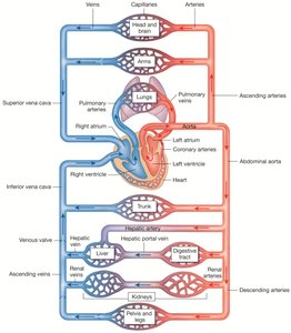
The cardiovascular system is essential for transporting materials throughout the body, including nutrients, gases, hormones, and waste products. It consists of the heart (the pump), blood (the fluid), and blood vessels (the tubes). Understanding the structure and function of this system is crucial for comprehending how the body maintains homeostasis and responds to physiological challenges.
Overview of the Cardiovascular System
Components and Functions
Heart: A muscular organ divided into left and right halves by the septum. Each half contains an atrium (receives blood) and a ventricle (pumps blood out).
Blood: Composed of cells (erythrocytes, leukocytes, platelets) and plasma.
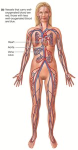
Blood Vessels: Arteries (carry blood away from the heart), veins (return blood to the heart), and capillaries (sites of exchange).
Pulmonary Circulation: Right side of the heart pumps deoxygenated blood to the lungs.
Systemic Circulation: Left side of the heart pumps oxygenated blood to the body.
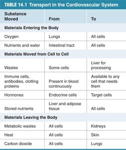
Transport Functions
Materials Entering the Body: Oxygen (lungs to all cells), nutrients and water (intestinal tract to all cells).
Materials Moved Between Cells: Hormones, immune cells, antibodies, and stored nutrients.
Materials Leaving the Body: Metabolic wastes (to kidneys), heat (to skin), carbon dioxide (to lungs).
Substance Moved | From | To |
|---|---|---|
Oxygen | Lungs | All cells |
Nutrients and water | Intestinal tract | All cells |
Wastes | Some cells | Liver for processing |
Immune cells, antibodies, clotting proteins | Present in blood continuously | Available to any cell that needs them |
Hormones | Endocrine cells | Target cells |
Stored nutrients | Liver and adipose tissue | All cells |
Metabolic wastes | All cells | Kidneys |
Heat | All cells | Skin |
Carbon dioxide | All cells | Lungs |
Blood Flow, Pressure, and Resistance
Principles of Fluid Flow
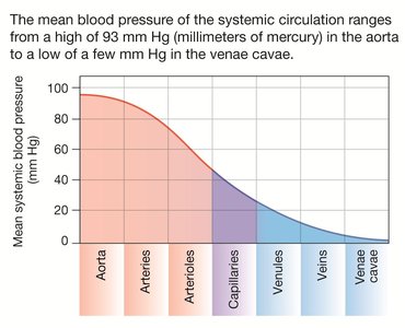
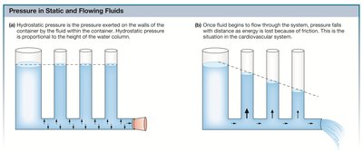
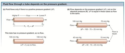
Blood flows from regions of higher pressure to regions of lower pressure. The heart creates pressure by contracting, and blood moves through vessels where pressure decreases due to friction and resistance.
Pressure Gradient (ΔP): The difference in pressure between two points drives flow.
Hydrostatic Pressure: Pressure exerted by a fluid at rest in all directions.
Flow Equation:
Resistance to Flow
Poiseuille’s Law: Resistance () is proportional to the length () of the vessel and the viscosity () of the fluid, and inversely proportional to the fourth power of the radius ():
Vasoconstriction: Decreases vessel radius, increasing resistance and decreasing flow.
Vasodilation: Increases vessel radius, decreasing resistance and increasing flow.
Velocity of Blood Flow
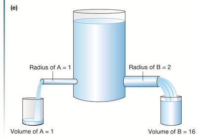
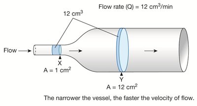
Flow Rate (Q): Volume of blood passing a point per unit time.
Velocity (v): Distance a volume of blood travels per unit time. , where is cross-sectional area.
Structure of the Heart
Anatomy and Chambers
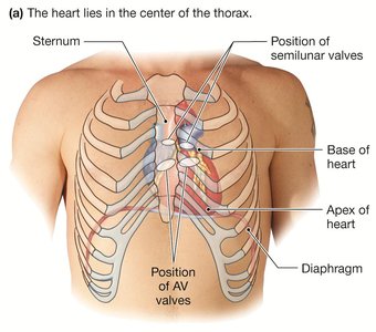
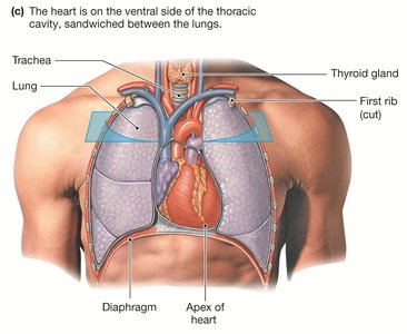
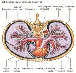
The heart is located in the center of the thorax, encased in the pericardium, and consists of four chambers: two atria and two ventricles.
The right side receives deoxygenated blood and pumps it to the lungs; the left side receives oxygenated blood and pumps it to the body.
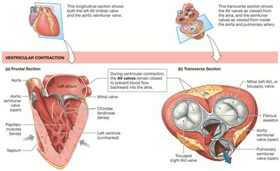
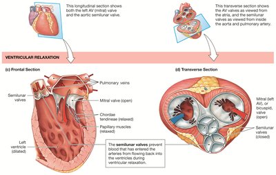
Heart Valves and Blood Flow
Atrioventricular (AV) Valves: Tricuspid (right) and bicuspid/mitral (left) valves prevent backflow from ventricles to atria.
Semilunar Valves: Aortic and pulmonary valves prevent backflow from arteries to ventricles.
Chordae Tendineae: Prevent valve eversion during ventricular contraction.
Coronary Circulation
The coronary arteries supply oxygenated blood to the heart muscle itself, while coronary veins return deoxygenated blood to the right atrium.
Cardiac Muscle and Electrical Activity
Cardiac Muscle Structure
Cardiac muscle cells are striated, branched, and connected by intercalated disks containing gap junctions for electrical coupling.
Autorhythmic (pacemaker) cells generate spontaneous action potentials, while contractile cells produce force.
Excitation-Contraction Coupling
Action potentials in pacemaker cells trigger Ca2+-induced Ca2+ release from the sarcoplasmic reticulum, leading to contraction.
Relaxation occurs as Ca2+ is pumped back into the SR or out of the cell via the Na+-Ca2+ exchanger.
Cardiac Action Potentials
Contractile Cells: Have a stable resting potential, rapid depolarization (Na+ influx), plateau phase (Ca2+ influx), and repolarization (K+ efflux).
Autorhythmic Cells: Unstable pacemaker potential, slow depolarization (If channels, Na+ influx), and Ca2+-dependent action potential.
Electrical Conduction and the Cardiac Cycle
Conduction System
SA node (pacemaker) initiates the heartbeat; signal spreads via internodal pathways to AV node, then through the bundle of His, bundle branches, and Purkinje fibers.
AV node delay allows atrial contraction before ventricular contraction.
Electrocardiogram (ECG/EKG)
Records the summed electrical activity of the heart.
P wave: Atrial depolarization
QRS complex: Ventricular depolarization (and atrial repolarization)
T wave: Ventricular repolarization
U wave: Repolarization of papillary muscles or Purkinje fibers (sometimes seen)
Mechanical Events of the Cardiac Cycle
Phases of the Cardiac Cycle
Atrial and Ventricular Diastole: Heart at rest, blood flows from veins to atria and ventricles.
Atrial Systole: Atria contract, completing ventricular filling (end-diastolic volume, EDV).
Isovolumic Ventricular Contraction: AV valves close (first heart sound, "lub"), pressure rises, no blood ejected.
Ventricular Ejection: Semilunar valves open, blood ejected into arteries (end-systolic volume, ESV).
Isovolumic Ventricular Relaxation: Semilunar valves close (second heart sound, "dup"), pressure falls, AV valves reopen.
Cardiac Output and Stroke Volume
Stroke Volume (SV):
Cardiac Output (CO):
Average CO at rest is about 5 L/min in adults.
Clinical Applications and Homeostatic Imbalances
Common Disorders
Broken Heart Syndrome (Takotsubo Cardiomyopathy): Temporary weakening of the heart muscle due to severe emotional or physical stress.
Pericarditis: Inflammation of the pericardium, causing chest pain and potential cardiac dysfunction.
Heart Murmurs: Abnormal heart sounds due to valve defects (incompetent or stenotic valves).
Arrhythmias: Irregular heart rhythms, including atrial and ventricular fibrillation.
Congestive Heart Failure (CHF): Inadequate cardiac output due to weakened myocardium, often resulting from coronary artery disease, hypertension, or myocardial infarction.
Key Equations and Concepts
Poiseuille’s Law:
Flow Equation:
Velocity of Flow:
Cardiac Output:
Additional info: This summary integrates foundational concepts in cardiovascular physiology, including structure, function, and clinical relevance, and is suitable for ANP college-level study.
