 Back
BackCardiovascular System: Anatomy and Blood Flow
Study Guide - Smart Notes
 Tailored notes based on your materials, expanded with key definitions, examples, and context.
Cardiovascular System: Structure and Function
Overview of the Cardiovascular and Circulatory Systems
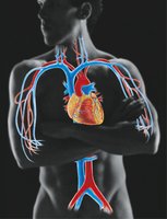
The cardiovascular system consists of the heart and blood vessels (arteries, veins, and capillaries). It is responsible for pumping blood throughout the body, delivering oxygen and nutrients to tissues, and removing waste products such as carbon dioxide. The circulatory system includes both the cardiovascular and lymphatic systems, working together to maintain homeostasis and fluid balance.
Heart: Muscular organ that pumps blood.
Blood vessels: Tubular structures (arteries, veins, capillaries) that transport blood.
Functions: Transport of gases, nutrients, hormones, and waste; regulation of temperature and pH; protection via immune cells.
Gross Anatomy of the Heart
Location, Size, and Orientation
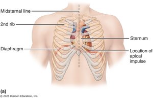
The heart is about the size of a fist and weighs less than 1 pound. It is located in the mediastinum, the medial cavity of the thorax, superior to the diaphragm. The heart is tilted obliquely, with two-thirds of its mass to the left of the midline and the apex pointing toward the left hip.
Mediastinum: Central compartment of the thoracic cavity.
Apex: Inferior, pointed end of the heart.
Base: Broad, superior portion where major vessels attach.
Coverings and Layers of the Heart Wall
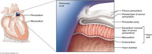
The heart is enclosed by the pericardium, which consists of two main layers:
Fibrous pericardium: Tough, outer layer that protects and anchors the heart.
Serous pericardium: Thin, double-layered membrane (parietal and visceral layers) with a lubricating pericardial cavity between them.
The heart wall has three layers:
Epicardium: Outer layer (visceral pericardium).
Myocardium: Middle, muscular layer responsible for contraction.
Endocardium: Inner layer of simple squamous epithelium lining the chambers and valves.
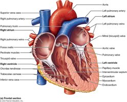
Chambers of the Heart and Associated Great Vessels
The heart has four chambers:
Atria (right and left): Superior, thin-walled receiving chambers.
Ventricles (right and left): Inferior, thick-walled pumping chambers.
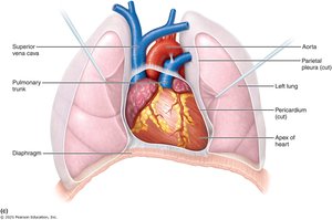
Major vessels associated with the heart:
Superior and inferior vena cava: Return deoxygenated blood to the right atrium.
Pulmonary trunk and arteries: Carry deoxygenated blood from the right ventricle to the lungs.
Pulmonary veins: Return oxygenated blood from the lungs to the left atrium.
Aorta: Delivers oxygenated blood from the left ventricle to the body.
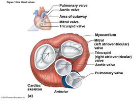
Heart Valves and Blood Flow
Types and Functions of Heart Valves
Heart valves ensure unidirectional blood flow through the heart. Their opening and closing are determined by pressure gradients across the valves.
Atrioventricular (AV) valves: Between atria and ventricles (tricuspid on right, bicuspid/mitral on left). Prevent backflow into atria.
Semilunar (SL) valves: Between ventricles and major arteries (pulmonary and aortic valves). Prevent backflow into ventricles.
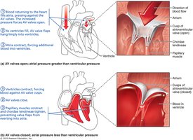
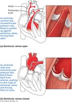
Operation of AV and SL Valves
AV valves open when atrial pressure exceeds ventricular pressure, allowing blood to flow into the ventricles. When ventricles contract, the increased pressure forces the valves shut, preventing backflow. SL valves open when ventricular pressure exceeds arterial pressure, allowing blood to exit the heart. When pressure falls, the valves close to prevent backflow.
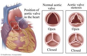
Heart Sounds and Valve Pathologies
The "lub-dup" sounds of the heart are caused by the closure of the AV valves (lub) and SL valves (dup). Valve disorders include:
Insufficient (incompetent) valve: Valve does not close properly, causing regurgitation.
Valvular stenosis: Valve does not open fully, restricting blood flow.
Both conditions can cause heart murmurs due to turbulent blood flow.
Blood Vessels: Structure and Function
Types of Blood Vessels
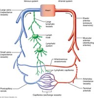
Blood vessels form a closed circuit for blood flow. The main types are:
Arteries: Carry blood away from the heart (usually oxygenated).
Arterioles: Small branches of arteries leading to capillaries.
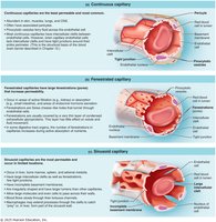
Capillaries: Microscopic vessels for exchange of gases, nutrients, and wastes.
Venules: Small vessels collecting blood from capillaries.
Veins: Carry blood toward the heart (usually deoxygenated).
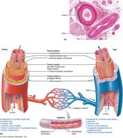
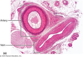
Structural Composition of Blood Vessel Walls
Blood vessel walls have three layers:
Tunica intima: Inner endothelial layer.
Tunica media: Middle layer of smooth muscle and elastic fibers (thicker in arteries).
Tunica externa (adventitia): Outer layer of connective tissue.
Types of Arteries, Capillaries, and Veins
Elastic arteries: Largest, near the heart, act as pressure reservoirs (e.g., aorta).
Muscular arteries: Medium-sized, distribute blood to organs.
Arterioles: Smallest arteries, regulate blood flow into capillaries.
Capillaries: Three types—continuous (least permeable), fenestrated (more permeable), and sinusoidal (most permeable).
Veins: Large lumens, thin walls, act as blood reservoirs, may contain valves.
Venules: Small veins collecting blood from capillaries.

Systemic and Pulmonary Circuits
Pathways of Blood Circulation
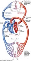
The heart pumps blood through two main circuits:
Systemic circuit: Left ventricle → aorta → body tissues (oxygen delivery) → vena cavae → right atrium.
Pulmonary circuit: Right ventricle → pulmonary arteries → lungs (gas exchange) → pulmonary veins → left atrium.
Special Circulations
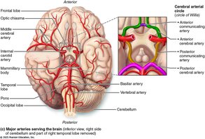
Circle of Willis
The Circle of Willis is a circular arterial structure at the base of the brain. It provides collateral circulation, stabilizing blood supply to the brain and offering alternate routes if major arteries are blocked.
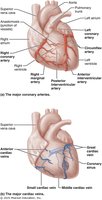
Coronary Circulation
Coronary circulation supplies the heart muscle (myocardium) with oxygen and nutrients. The coronary arteries branch from the aorta, and the coronary sinus returns deoxygenated blood to the right atrium. Pathologies include angina pectoris, myocardial infarction (heart attack), and coronary artery disease (atherosclerosis).
Path of Blood Through the Heart
Stepwise Pathway
Blood flows through the heart in the following sequence:
Right atrium (RA)
Tricuspid valve (Tri)
Right ventricle (RV)
Pulmonary semilunar valve (PSLV)
Pulmonary trunk
Right and left pulmonary arteries
Lungs (gas exchange)
Right and left pulmonary veins
Left atrium (LA)
Bicuspid (mitral) valve (Bi)
Left ventricle (LV)
Aortic semilunar valve (ASLV)
Aorta
Body tissues (systemic circulation)
Superior/inferior vena cava (S/I Vena Cava)
Back to right atrium
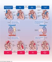
Additional info: The color coding (red for oxygen-rich, blue for oxygen-poor) is a standard convention in cardiovascular diagrams. The sequence above highlights the unidirectional flow and the separation of oxygenated and deoxygenated blood in the heart and vessels.
