 Back
BackCell Communication, Endocrine System, Blood, and Cardiovascular System Study Notes
Study Guide - Smart Notes
 Tailored notes based on your materials, expanded with key definitions, examples, and context.
How Cells Communicate
Types of Cell Communication
Cells communicate through various mechanisms to coordinate physiological processes. These include direct, synaptic, autocrine, paracrine, and endocrine communication.
Direct Communication: Cells communicate through gap junctions, allowing ions and small molecules to pass directly between adjacent cells.
Synaptic Communication: Neurons release neurotransmitters across synapses to target cells, enabling rapid and specific signaling.
Autocrine Communication: Cells release signals that bind to receptors on their own surface, affecting themselves.
Paracrine Communication: Signals are released to affect nearby cells within the same tissue.
Endocrine Communication: Endocrine cells release hormones into the bloodstream, which travel to distant target organs.
Receptor Types and Signal Transduction
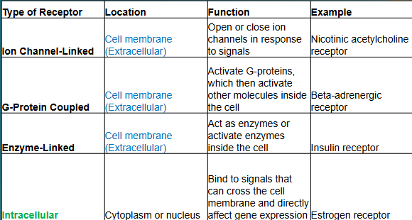
Cell signaling depends on the type and location of receptors. Receptors can be classified based on their location and mechanism of action.
Type of Receptor | Location | Function | Example |
|---|---|---|---|
Ion Channel-Linked | Cell membrane (Extracellular) | Open or close ion channels in response to signals | Nicotinic acetylcholine receptor |
G-Protein Coupled | Cell membrane (Extracellular) | Activate G-proteins, which then activate other molecules inside the cell | Beta-adrenergic receptor |
Enzyme-Linked | Cell membrane (Extracellular) | Act as enzymes or activate enzymes inside the cell | Insulin receptor |
Intracellular | Cytoplasm or nucleus | Bind to signals that can cross the cell membrane and directly affect gene expression | Estrogen receptor |
The Endocrine System
Major Endocrine Glands and Hormones
The endocrine system consists of glands that secrete hormones to regulate body functions. The hypothalamus controls the pituitary gland, which in turn regulates other endocrine glands.
Pituitary Gland: Divided into anterior (adenohypophysis) and posterior (neurohypophysis) lobes.
Hypothalamus: Regulates the pituitary gland and integrates nervous and endocrine functions.
Thyroid Gland: Releases thyroid hormones (regulate metabolism) and calcitonin (regulates calcium).
Parathyroid Glands: Release parathyroid hormone (regulates calcium).
Pancreas: Releases insulin (lowers blood glucose) and glucagon (raises blood glucose).
Adrenal Glands: Cortex releases cortisol, mineralocorticoids, and androgens; medulla releases epinephrine.
Pituitary Hormones
Anterior Pituitary (Adenohypophysis): Releases six hormones via blood vessels from the hypothalamus:
Growth Hormone (GH): Stimulates bone and tissue growth.
Adrenocorticotropic Hormone (ACTH): Stimulates adrenal cortex to release cortisol.
Thyroid Stimulating Hormone (TSH): Stimulates thyroid gland.
Gonadotropic Hormones (FSH/LH): Regulate reproductive functions.
Prolactin (PRL): Stimulates milk production.
Posterior Pituitary (Neurohypophysis): Releases two hormones via nerve fibers from the hypothalamus:
Antidiuretic Hormone (ADH): Signals kidneys to conserve water.
Oxytocin (OT): Triggers uterine contractions and milk let-down.
Blood: Composition and Function
Components of Blood
Blood is a connective tissue composed of plasma and formed elements. It transports gases, nutrients, hormones, and waste products throughout the body.
Plasma (55%): Water, ions, nutrients, proteins, gases, and waste products.
Formed Elements (45%):
Red Blood Cells (Erythrocytes): Carry oxygen via hemoglobin, biconcave shape, no nucleus, lifespan ~120 days.
White Blood Cells (Leukocytes): Immune defense, classified as granular or agranular.
Platelets (Thrombocytes): Involved in blood clotting.
Leukocytes (White Blood Cells)
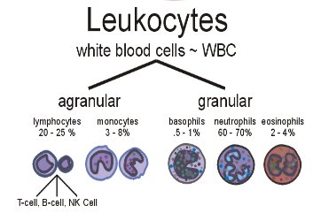
Leukocytes are divided into two main groups based on the presence or absence of granules in their cytoplasm.
Agranular Leukocytes: Lymphocytes (20-25%), Monocytes (3-8%)
Granular Leukocytes: Basophils (0.5-1%), Neutrophils (60-70%), Eosinophils (2-4%)
Granular Leukocytes
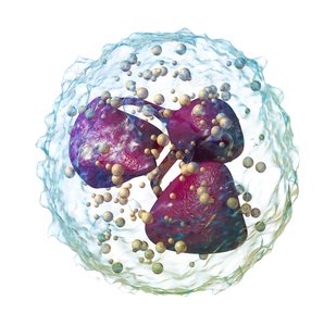
Neutrophils: Most common WBC, polymorphonuclear, first responders to infection, phagocytize bacteria and fungi.
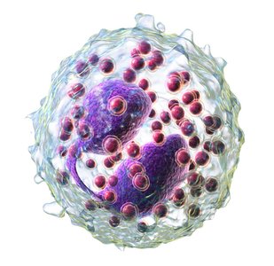
Eosinophils: Attack parasitic worms, involved in allergic responses, control inflammation with enzymes.
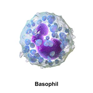
Basophils: Rarest WBC, release histamine (vasodilation) and heparin (prevents clotting).
Agranular Leukocytes
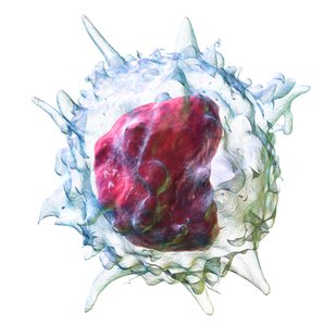
Monocytes: Largest leukocyte, differentiate into macrophages in tissues, phagocytize pathogens and debris, secrete substances to attract immune cells and fibroblasts.
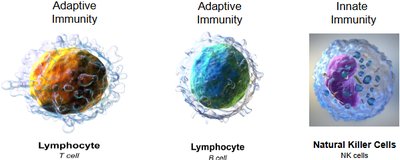
Lymphocytes: Key cells in adaptive immunity (T cells, B cells) and innate immunity (Natural Killer cells).
Blood Vessels and Circulation
Structure of Blood Vessels
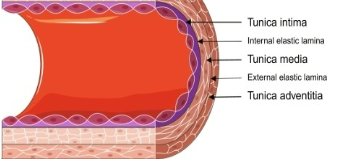
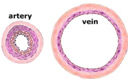
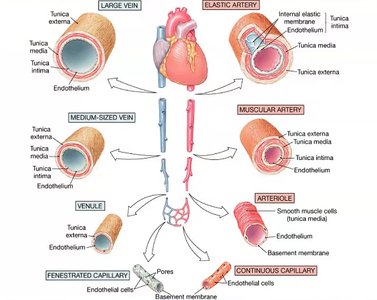
Blood vessels have three main layers (tunics) that vary in thickness and composition depending on the vessel type.
Tunica Intima: Innermost layer, simple squamous epithelium, connective tissue, elastic fibers.
Tunica Media: Middle layer, smooth muscle, external elastic membrane; thick in arteries, thin in veins.
Tunica Externa (Adventitia): Outermost layer, fibrous connective tissue, provides protection.
Arteries vs. Veins
Arteries: Thick tunica media, maintain high pressure, smaller lumen, no valves.
Veins: Thinner tunica media, larger lumen, contain valves to prevent backflow.
Types of Capillaries
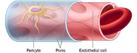
Capillaries are the smallest blood vessels and are specialized for exchange between blood and tissues.
Continuous Capillaries: Permit diffusion of water and small solutes; most common type.
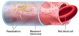
Fenestrated Capillaries: Have pores for passage of larger molecules; found in kidneys, intestines, endocrine glands.
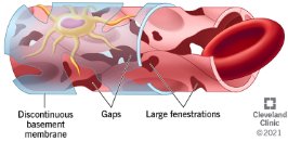
Sinusoids: Large gaps and discontinuous basement membrane; allow passage of proteins and cells; found in liver, spleen, bone marrow.
Major Circulatory Pathways
Pulmonary Circuit: Carries deoxygenated blood from the heart to the lungs and returns oxygenated blood to the heart.
Systemic Circuit: Delivers oxygenated blood from the heart to the body and returns deoxygenated blood to the heart.
Arteries: Carry blood away from the heart.
Veins: Carry blood toward the heart.
Capillaries: Connect arteries and veins; site of exchange.
Special Circulatory Pathways
Cerebral Arterial Circle (Circle of Willis)
A circulatory anastomosis that supplies blood to the brain and surrounding structures.
Hepatic Portal System
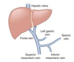
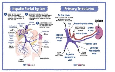
The hepatic portal system directs blood from parts of the gastrointestinal tract to the liver for processing and detoxification before it enters the systemic circulation.
Fetal Circulation
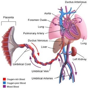
Fetal circulation includes unique structures that allow blood to bypass the nonfunctional fetal lungs and liver.
Umbilical Vein: Carries oxygenated blood from placenta to fetus.
Umbilical Arteries: Carry deoxygenated blood from fetus to placenta.
Placenta: Site of exchange between maternal and fetal blood (no mixing of blood).
The Heart
Pericardial Membranes and Heart Wall Layers
Pericardium: Double-walled sac enclosing the heart.
Pericardial Sac: Outer fibrous layer and inner serous layer.
Parietal Pericardium: Outer layer of serous pericardium.
Visceral Pericardium (Epicardium): Inner layer, covers the heart surface.
Serous Fluid: Reduces friction between layers.
Myocardium: Muscular layer responsible for contraction.
Cardiac Tissue Characteristics
Smaller cells, 1-5 nuclei per cell.
Intercalated discs for cell adhesion and electrical signal transmission.
Abundant mitochondria and blood supply.
Capable of generating action potentials.
Endocardium lines internal chambers.
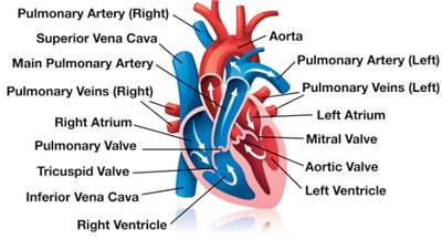
Direction of Blood Flow Through the Heart
Blood flows through the heart in a specific sequence, ensuring efficient oxygenation and distribution throughout the body.
Impulse Conduction System of the Heart
Sinoatrial (SA) Node: Pacemaker, initiates action potential, located in right atrium wall.
Internodal Pathways: Conduct impulses from SA node to AV node.
Atrioventricular (AV) Node: Located in floor of right atrium, slows impulse, allows atrial contraction.
Bundle of His: Conducts impulse from AV node to ventricles.
Bundle Branches: Carry impulse through interventricular septum.
Purkinje Fibers: Distribute impulse through ventricular myocardium, trigger ventricular contraction.
Pulse Points
The pulse is the palpable beat of the heart felt at various arterial sites, reflecting ventricular contraction.
Temporal (head)
Carotid (neck)
Apical (heart)
Brachial (arm)
Radial (wrist)
Femoral (thigh)
Popliteal (back of knee)
Posterior tibial (ankle)
Dorsalis pedis (foot)
