 Back
BackCell Communication, Integration, and Homeostasis: Chapter 6 Study Notes
Study Guide - Smart Notes
 Tailored notes based on your materials, expanded with key definitions, examples, and context.
Communication, Integration, and Homeostasis
Introduction to Cell Communication
Cell communication is essential for maintaining physiological balance and coordinating functions across tissues and organs. It enables cells to respond to internal and external signals, integrate information, and maintain homeostasis.
Homeostasis: The process by which organisms maintain a stable internal environment.
Integration: The coordination of multiple cellular responses to achieve a unified physiological outcome.
Response: The cellular change triggered by a signal, such as altering protein activity or gene expression.
6.1 Cell-to-Cell Communication: Overview
Physiological Signals
Cells communicate using electrical and chemical signals. Electrical signals involve changes in membrane potential, while chemical signals are secreted into the extracellular fluid (ECF) and bind to target cells.
Electrical signals: Changes in a cell's membrane potential.
Chemical signals: Molecules secreted by cells that bind to receptors on target cells.
Target cells: Cells that respond to electrical or chemical signals.
Four Basic Methods of Cell Communication
Cells use four primary methods to communicate, each with distinct mechanisms and ranges.
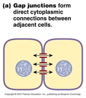
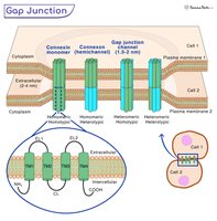
Gap junctions: Direct cytoplasmic connections between adjacent cells, allowing ions and small molecules to pass freely.
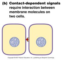
Contact-dependent signals: Require interaction between membrane molecules on two cells.
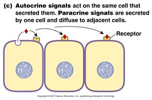
Autocrine signals: Act on the same cell that secreted them.
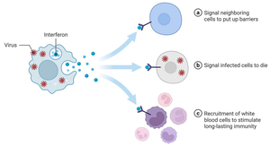
Paracrine signals: Secreted by one cell and diffuse to adjacent cells.
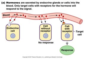
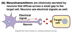
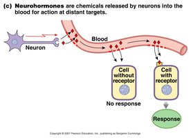
Long-distance communication: Involves hormones (endocrine signaling) and neurocrine molecules (neurotransmitters and neurohormones).
6.2 Signal Pathways: Overview
General Steps in Signal Pathways
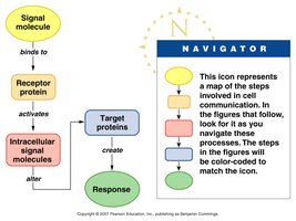
All signal pathways share a common sequence of events:
Ligand binds to a protein receptor.
Ligand-receptor binding activates the receptor.
Receptor activates one or more intracellular signal molecules.
Last signal molecule creates a response by modifying existing proteins or initiating synthesis of new proteins.
Receptor Locations
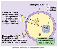
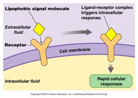
Receptors can be located in the cytosol, nucleus, or on the cell membrane. Lipophilic (fat-soluble) signals diffuse through the membrane, while lipophobic (water-soluble) signals bind to surface receptors.
Lipophilic signals: Bind to cytosolic or nuclear receptors; slower responses.
Lipophobic signals: Bind to membrane receptors; rapid responses.
Categories of Membrane Receptors
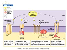
There are four main categories of membrane receptors:
Ligand-gated channels: Open or close in response to ligand binding.
Receptor enzymes: Activate intracellular enzymes.
G protein-coupled receptors (GPCR): Activate G proteins, which regulate ion channels or enzymes.
Integrin receptors: Link the cytoskeleton to the extracellular matrix.
Signal Transduction
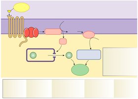
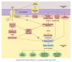
Signal transduction is the transmission of information from one side of a membrane to the other using membrane proteins. It involves first messengers (extracellular signals), second messengers (intracellular signals), and a cellular response.
First messenger: The extracellular signal molecule.
Second messenger: Intracellular molecules that relay the signal.
Response: The final cellular effect.
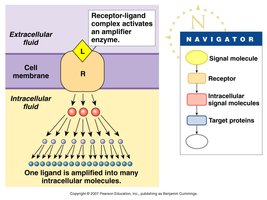
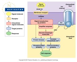
Basic Transduction Pathway
Membrane receptors and associated proteins activate protein kinases, amplifier enzymes, alter ion channel gating, or increase calcium binding to proteins.
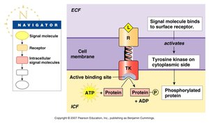
Protein kinases: Enzymes that transfer phosphate groups from ATP to proteins.
Amplifier enzymes: Create intracellular second messengers.
Ion channel gating: Changes membrane permeability.
Calcium binding: Alters protein activity.
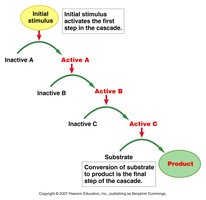
Signal Transduction Cascades
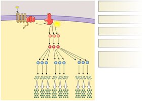
Signal transduction often forms cascades, where an initial stimulus activates a series of steps, amplifying the response.
Cascade: Sequential activation of molecules, leading to a final product.
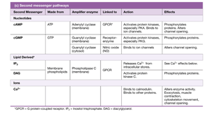
Second Messenger Pathways
Second messengers relay signals inside the cell. Common second messengers include cAMP, cGMP, IP3, DAG, and Ca2+.
Second Messenger | Made From | Amplifier Enzyme | Linked to | Action | Effects |
|---|---|---|---|---|---|
cAMP | ATP | Adenylyl cyclase | GPCR | Activates protein kinases, binds to ion channels | Phosphorylates proteins, alters channel opening |
cGMP | GTP | Guanylyl cyclase | Receptor enzyme, GPCR | Activates protein kinases, binds to ion channels | Phosphorylates proteins, alters channel opening |
IP3 | Membrane phospholipids | Phospholipase C | GPCR | Releases Ca2+ from intracellular stores | See Ca2+ effects |
DAG | Membrane phospholipids | Phospholipase C | GPCR | Activates protein kinase C | Phosphorylates proteins |
Ca2+ | Ion | None | Channel | Binds to calmodulin, other proteins | Alters enzyme activity, exocytosis, movement, channel opening |
Membrane Receptor Types
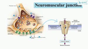
Receptor-channel: Opens or closes in response to signal molecule binding.
G protein-coupled receptor (GPCR): Activates G proteins, which open ion channels or alter enzyme activity.
Receptor enzyme: Activates intracellular enzymes, such as tyrosine kinase.
Summary of Signal Transduction Systems
6.3 Novel Signal Molecules
Calcium as a Signal Molecule
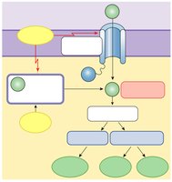
Calcium ions (Ca2+) act as important intracellular messengers, binding to proteins and altering their activity. Calcium signaling is involved in exocytosis, movement, and enzyme activation.
Gaseous Signal Molecules
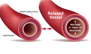
Gases such as nitric oxide (NO) serve as signaling molecules. NO acts as a neurotransmitter and neuromodulator, is produced by endothelial cells, and causes vasodilation by relaxing smooth muscle in blood vessels.
Lipid Signal Molecules
Lipid-derived messengers, such as those produced by the arachidonic acid cascade, include leukotrienes and prostanoids (prostaglandins, thromboxanes). These molecules play roles in inflammation, pain, fever, and allergic responses.
Leukotrienes: Involved in asthma and anaphylaxis.
Prostaglandins: Regulate sleep, inflammation, pain, and fever.
Thromboxanes: Involved in blood clotting.
NSAIDs: Inhibit COX enzymes to reduce inflammation.
6.4 Modulation of Signal Pathways
Specificity and Competition
Receptors exhibit specificity and competition. Agonists activate receptors, while antagonists block them. Multiple receptors may exist for one ligand, leading to different responses (e.g., alpha and beta adrenergic receptors).
Agonist: Molecule that activates a receptor.
Antagonist: Molecule that blocks a receptor.
Alpha receptor: Causes vasoconstriction.
Beta receptor: Causes vasodilation.
Up-Regulation vs. Down-Regulation
Cells can increase (up-regulate) or decrease (down-regulate) the number of receptors in response to changes in signal concentration.
Up-regulation: Increase in receptor number, enhancing sensitivity.
Down-regulation: Decrease in receptor number, reducing sensitivity.
6.5 Control Pathways: Feedback Loops
Feedback Loops in Homeostasis
Feedback loops are essential for maintaining homeostasis. Negative feedback loops counteract changes, while positive feedback loops amplify them.
Negative feedback: Reduces the effect of a stimulus, restoring balance.
Positive feedback: Enhances the effect of a stimulus, leading to a greater response.
