 Back
BackCell Cycle and Protein Synthesis: Structure and Function in Human Anatomy & Physiology
Study Guide - Smart Notes
 Tailored notes based on your materials, expanded with key definitions, examples, and context.
Cells: The Living Units
Overview of the Cell Cycle
The cell cycle is a series of events that cells undergo from the time they are formed until they divide into two daughter cells. It is essential for growth, repair, and maintenance in multicellular organisms. The cell cycle consists of two major periods: interphase and cell division (mitotic phase).
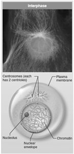
Interphase: The cell grows, carries out normal functions, and prepares for division. The nuclear material is in the form of uncondensed chromatin.
Cell Division (Mitotic Phase): The cell divides into two daughter cells, ensuring genetic continuity.
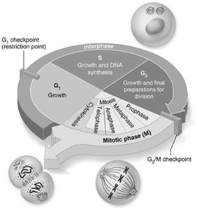
Interphase
Interphase is the period when the cell performs its normal metabolic activities and prepares for division. It is subdivided into three subphases:
G1 (Gap 1): Vigorous growth and metabolism occur. Cells that permanently cease dividing enter the G0 phase.
S (Synthesis): DNA replication takes place, ensuring each daughter cell receives an identical set of genetic material.
G2 (Gap 2): Final preparations for cell division are made.
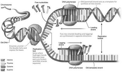
DNA Replication
Before a cell divides, it must replicate its DNA so that each daughter cell receives a complete set of genetic instructions. The process involves:
Unwinding and unzipping of the double-stranded DNA helix by enzymes.
Formation of a replication fork (point where strands separate) and a replication bubble (active area of replication).
RNA primers initiate synthesis; DNA polymerase adds nucleotides to form new strands (one leading, one lagging).
DNA ligase joins short segments of the lagging strand.
The result is two identical "daughter" DNA molecules, each with one old and one new strand (semiconservative replication).
Cell Division: Mitosis and Cytokinesis
Most cells divide regularly for growth and repair, except for skeletal, cardiac, and nerve cells. Cell division consists of mitosis (nuclear division) and cytokinesis (cytoplasmic division).
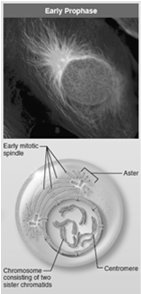
Mitosis: Ensures each daughter cell receives a full set of chromosomes. It occurs in four stages:
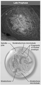
Prophase: Chromatin condenses into chromosomes; spindle fibers form; nuclear envelope breaks down.
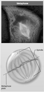
Metaphase: Chromosomes align at the cell's equator (metaphase plate).
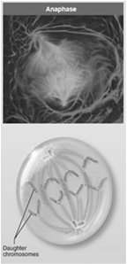
Anaphase: Sister chromatids separate and move toward opposite poles.
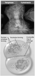
Telophase: Chromosomes decondense; nuclear envelopes reform; nucleoli reappear.
Cytokinesis: Division of the cytoplasm, resulting in two separate daughter cells.
Control of Cell Division
Cell division is tightly regulated by "go" and "stop" signals:
Go signals: Include critical surface-to-volume ratio and chemical factors (e.g., growth factors, hormones).
Stop signals: Include contact inhibition (cells stop dividing when in contact with other cells) and lack of space.
Protein Synthesis
Genetic Code and the Role of DNA
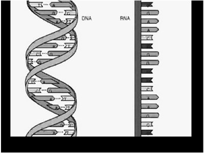
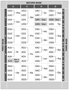
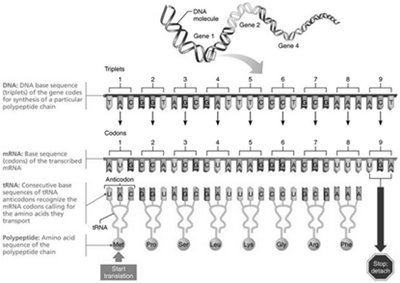
DNA serves as the master blueprint for protein synthesis, determining the order of amino acids in a polypeptide. A gene is a segment of DNA that codes for one polypeptide. The genetic code is based on the sequence of nitrogenous bases (adenine, guanine, cytosine, thymine) in triplets, each specifying a particular amino acid.
The Role of RNA
RNA acts as the intermediary between DNA and protein synthesis. It is synthesized in the nucleus and differs from DNA by having uracil instead of thymine and ribose instead of deoxyribose. There are three main types of RNA:
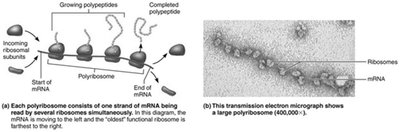
Messenger RNA (mRNA): Carries the genetic code from DNA to ribosomes; produced by transcription.
Ribosomal RNA (rRNA): Structural component of ribosomes, facilitating protein synthesis.
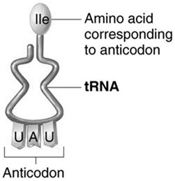
Transfer RNA (tRNA): Brings specific amino acids to the ribosome during translation; contains an anticodon complementary to mRNA codons.
Steps of Protein Synthesis
Transcription: DNA information is coded into mRNA in the nucleus.
Translation: mRNA is decoded at the ribosome to assemble a polypeptide chain.
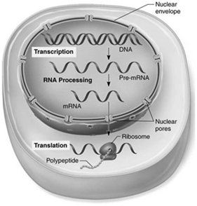
Transcription
Transcription occurs in three phases:
Initiation: RNA polymerase binds to DNA and separates the strands.
Elongation: RNA polymerase adds complementary RNA nucleotides.
Termination: Transcription ends when a termination signal is reached.
Translation
Translation is the process of converting the nucleotide sequence of mRNA into an amino acid sequence of a protein. It involves:
Initiation: Ribosome assembles around the start codon (AUG) on mRNA and the initiator tRNA (methionine).
Elongation: tRNAs bring amino acids to the ribosome, matching their anticodons to mRNA codons. Peptide bonds form between amino acids.
Termination: When a stop codon is reached, the polypeptide is released.
Summary Table: Types of RNA and Their Functions
Type of RNA | Function |
|---|---|
mRNA (Messenger RNA) | Carries genetic code from DNA to ribosome for translation |
rRNA (Ribosomal RNA) | Forms the core of ribosome's structure and catalyzes protein synthesis |
tRNA (Transfer RNA) | Brings specific amino acids to the ribosome during translation |
Cell Differentiation, Death, and Growth
Cell Differentiation
Although all cells contain the same DNA, they differentiate into various types with specialized functions due to selective gene expression. This process is regulated by chemical signals during development.
Cell Death and Growth
Apoptosis: Programmed cell death, important for removing damaged, infected, or unnecessary cells.
Hyperplasia: Accelerated cell growth, increasing cell numbers when needed.
Atrophy: Decrease in cell size due to loss of stimulation or use.
Cell division continues throughout life for tissue growth and repair, but is tightly regulated to maintain tissue integrity.
