 Back
BackCell Membrane Transport and Epithelial Transport: Mechanisms and Clinical Relevance
Study Guide - Smart Notes
 Tailored notes based on your materials, expanded with key definitions, examples, and context.
Cell Membrane Transport: Principles and Mechanisms
Introduction to Membrane Transport
Transport across cell membranes is essential for maintaining cellular homeostasis, nutrient uptake, waste removal, and signal transduction. The direction and rate of transport are governed by energy gradients and the properties of the transported molecules and the membrane itself.
Driving Forces for Membrane Transport
Chemical, Electrical, and Electrochemical Driving Forces
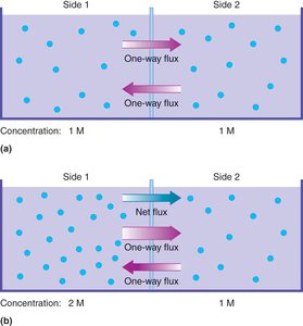
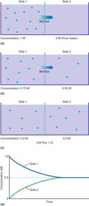
Chemical Driving Force: Arises from concentration gradients; molecules move from high to low concentration.
Electrical Driving Force: Due to membrane potential; cations are attracted to negative membrane potential, anions are repelled.
Electrochemical Driving Force: The sum of chemical and electrical forces; determines the net direction of ion movement.
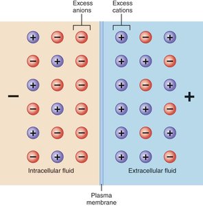
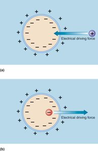
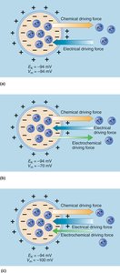
Equilibrium Potentials and the Nernst Equation
The Nernst equation calculates the equilibrium potential for an ion, where the electrical and chemical driving forces are balanced.
At equilibrium potential, there is no net movement of the ion across the membrane.
Types of Membrane Transport
Passive Transport
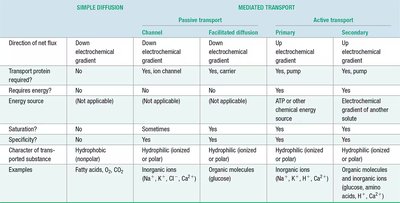
Simple Diffusion: Movement of molecules directly through the lipid bilayer, driven by concentration gradients. Only small, nonpolar molecules (e.g., O2, CO2, fatty acids) diffuse this way.
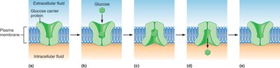
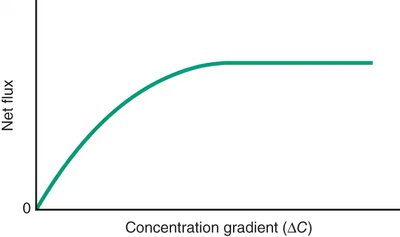
Facilitated Diffusion: Utilizes carrier proteins or channels for hydrophilic molecules (e.g., glucose, ions). Movement is still down the electrochemical gradient.
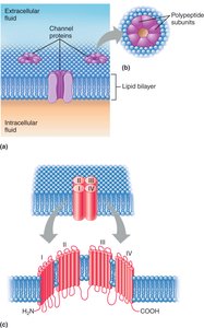
Diffusion Through Channels: Ion channels and aquaporins allow specific ions or water to cross membranes rapidly.
Active Transport
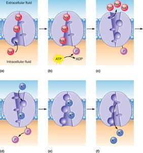
Primary Active Transport: Direct use of ATP to move substances against their electrochemical gradients (e.g., Na+/K+ pump).
Secondary Active Transport: Uses the energy stored in gradients created by primary active transport to move other substances (e.g., sodium-glucose cotransport).
Epithelial Transport: Structure and Function
Epithelial Structure and Polarity
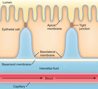
Epithelial tissues line body surfaces and cavities, forming barriers and regulating transport between compartments. Epithelial cells are polarized, with distinct apical (lumen-facing) and basolateral (blood/interstitial fluid-facing) membranes, each with unique transport proteins. Tight junctions between cells restrict paracellular movement and maintain compartmentalization.
Mechanisms of Epithelial Solute Transport
Transcellular Transport: Movement of substances through the cell, involving both apical and basolateral membranes.
Paracellular Transport: Movement between cells, limited by tight junctions.
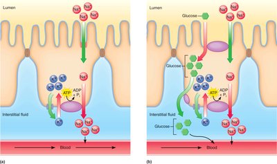
Example: In the intestine, Na+ is absorbed via apical channels and actively pumped out basolaterally; glucose is co-transported with Na+ and exits via facilitated diffusion.
Epithelial Water Transport
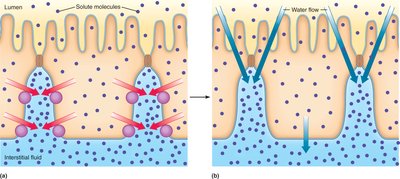
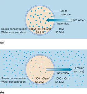
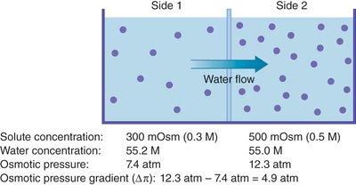
Water follows solute movement by osmosis, moving from areas of low to high solute concentration (or up the osmotic pressure gradient).
Active solute transport creates osmotic gradients that drive water absorption or secretion.
Clinical Connection: Cystic Fibrosis
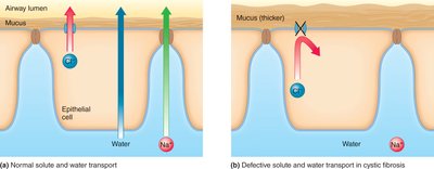
Cystic fibrosis is caused by defective chloride channels in epithelial cells, impairing Cl- and Na+ transport, reducing water secretion, and resulting in thick, sticky mucus in the airways.
Transcytosis
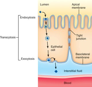
Large molecules cross epithelia via transcytosis, which combines endocytosis (uptake) and exocytosis (release) without lysosomal degradation.
Osmosis and Cell Volume Regulation
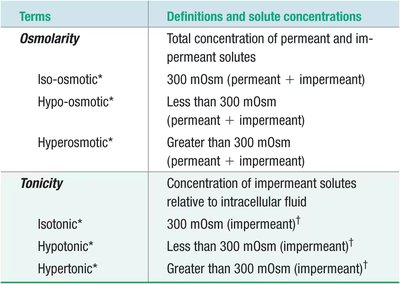
Osmosis and Osmolarity
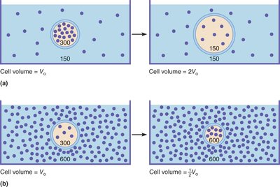
Osmosis: Passive movement of water across membranes, driven by differences in water concentration (or solute concentration).
Osmolarity: Total solute particle concentration of a solution (osmoles/L).
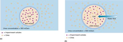
Tonicity: Effect of a solution on cell volume, determined by the concentration of nonpermeant solutes.
Vesicular Transport: Endocytosis, Exocytosis, and Transcytosis
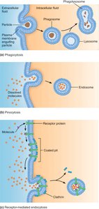
Endocytosis
Phagocytosis: "Cell eating"; uptake of large particles by membrane extensions.
Pinocytosis: "Cell drinking"; nonspecific uptake of extracellular fluid and solutes.
Receptor-mediated Endocytosis: Specific uptake of molecules via receptor binding and vesicle formation.
Exocytosis
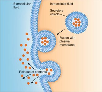
Vesicles fuse with the plasma membrane to release contents outside the cell; important for secretion, membrane recycling, and receptor recycling.
Transcytosis
Combination of endocytosis and exocytosis for transporting macromolecules across cells, especially in epithelial barriers.
Summary Table: Characteristics of Transport Mechanisms
Key Equations
van’t Hoff’s Equation for Free Energy Change:
Nernst Equation for Equilibrium Potential:
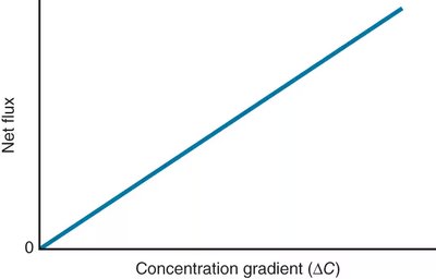
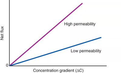
Fick’s Law for Simple Diffusion: where = flux, = permeability, = surface area, = concentration gradient
Osmotic Pressure: where = osmotic pressure, = solute concentration, = gas constant, = temperature
Clinical Applications and Examples
Cystic Fibrosis: Defective chloride channels impair water secretion, leading to thick mucus and respiratory complications.
Diabetes Mellitus: Impaired glucose transport affects cell energy and blood glucose levels, with consequences for osmolarity and cell volume.
IV Solutions: Isotonic, hypotonic, and hypertonic solutions are used clinically to manage patient fluid balance and cell volume.
