 Back
BackCell Membranes and Body Fluid Compartments: Structure, Composition, and Dynamics
Study Guide - Smart Notes
 Tailored notes based on your materials, expanded with key definitions, examples, and context.
Chapter 3: Compartmentation – Cells and Tissues
Anatomical and Functional Body Compartments
The human body is organized into anatomical cavities and functional fluid compartments, each separated by membranes that regulate the movement of substances.
Anatomical Compartments: Cranial, thoracic, and abdominopelvic cavities house major organs and are separated by physical barriers such as the diaphragm.
Functional Compartments: Body fluids are divided into intracellular fluid (ICF) and extracellular fluid (ECF). The ECF is further subdivided into interstitial fluid (surrounding cells) and blood plasma (within blood vessels).
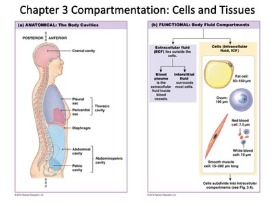
Example: The cranial cavity contains the brain, while the thoracic cavity contains the heart and lungs. The ICF is the fluid inside cells, and the ECF surrounds them.
Chapter 3: Cell Membrane Structure and Composition
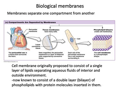
Biological Membranes
Biological membranes separate different compartments within the body and within cells, maintaining distinct environments necessary for physiological function.
Cell Membrane: Originally thought to be a single lipid layer, it is now known to be a phospholipid bilayer with embedded proteins.
Function: Controls the movement of substances into and out of cells, maintaining homeostasis.
Cell Membrane Lipids
The cell membrane is primarily composed of three types of lipids: phospholipids, sphingolipids, and cholesterol. These molecules contribute to the membrane's structure and function.
Phospholipids: Amphipathic molecules with hydrophilic heads and hydrophobic tails, forming bilayers in aqueous environments.
Sphingolipids: Often associated with lipid-anchored proteins and cell signaling.
Cholesterol: Adds flexibility and decreases permeability to small water-soluble molecules.
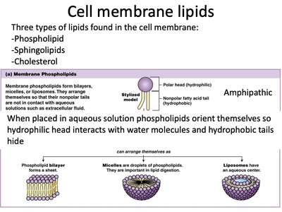
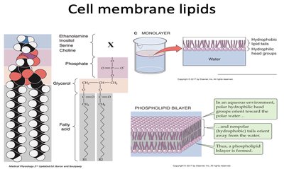
Example: In water, phospholipids spontaneously form bilayers, micelles, or liposomes, with hydrophobic tails hidden from water.
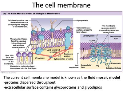
The Fluid Mosaic Model
The current model of the cell membrane is the fluid mosaic model, which describes a dynamic structure with proteins and lipids moving laterally within the bilayer.
Proteins: Dispersed throughout the membrane, serving various functions such as transport, signaling, and structural support.
Carbohydrates: Present as glycoproteins and glycolipids on the extracellular surface, involved in cell recognition and communication.
Membrane Composition
Cell membranes vary in their composition of proteins, lipids, and carbohydrates, depending on their function and metabolic activity.
Membrane | Protein | Lipid | Carbohydrate |
|---|---|---|---|
Red blood cell membrane | 49% | 43% | 8% |
Myelin membrane (nerve cells) | 18% | 79% | 3% |
Inner mitochondrial membrane | 76% | 24% | 0% |
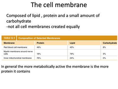
Key Point: More metabolically active membranes contain a higher proportion of proteins.
Chapter 3: Cell Membrane Proteins
Types and Roles of Membrane Proteins
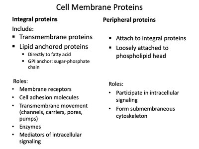
Membrane proteins can be classified as integral or peripheral based on their association with the lipid bilayer.
Integral Proteins: Span the membrane (transmembrane) or are anchored via lipids. Functions include transport, signaling, and cell adhesion.
Peripheral Proteins: Loosely attached to the membrane or integral proteins, involved in intracellular signaling and cytoskeletal attachment.
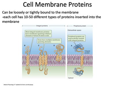
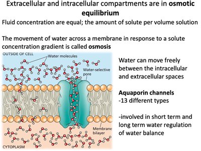
Example: Aquaporins (water channels) are integral proteins that facilitate water movement across the membrane.
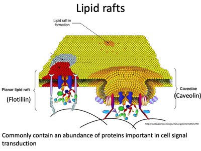
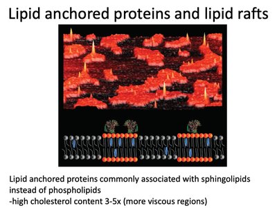
Lipid Rafts and Anchored Proteins
Lipid rafts are specialized membrane microdomains rich in cholesterol and sphingolipids, often involved in cell signaling.
Lipid-anchored proteins: Commonly associated with sphingolipids and have high cholesterol content, making these regions more viscous.
Function: Concentrate signaling molecules and facilitate communication within the cell.
Chapter 3: Cell Membrane Carbohydrates
Glycoproteins and Glycolipids
Carbohydrates are present on the extracellular surface of the cell membrane, attached to proteins (glycoproteins) or lipids (glycolipids).
Glycoproteins: Involved in cell recognition, signaling, and immune responses.
Glycolipids: Serve as recognition sites for cell-cell interactions.
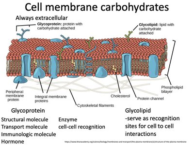
Example: Blood group antigens are glycoproteins and glycolipids on red blood cell membranes.
Chapter 5: Membrane Dynamics
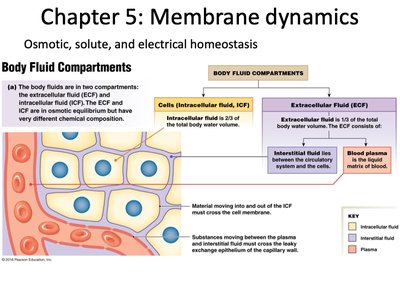
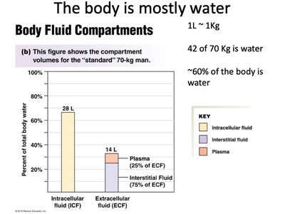
Body Fluid Compartments and Homeostasis
Body fluids are distributed between intracellular and extracellular compartments, each with distinct compositions and roles in maintaining homeostasis.
Intracellular Fluid (ICF): Makes up about two-thirds of total body water.
Extracellular Fluid (ECF): Includes interstitial fluid and plasma, making up the remaining one-third.
Osmotic, Chemical, and Electrical Equilibrium
Although water moves freely between compartments (osmotic equilibrium), the distribution of solutes (ions) creates chemical and electrical disequilibrium.
Osmosis: Movement of water across a membrane in response to solute concentration gradients.
Aquaporin Channels: Specialized proteins that facilitate water movement.
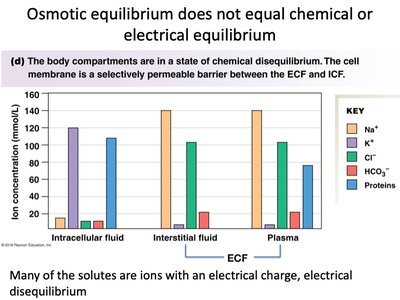
Key Point: The cell membrane is selectively permeable, allowing water but restricting many solutes, thus maintaining distinct internal environments.
Comparing Osmolarities
Osmolarity is a quantitative measure of solute concentration, while tonicity describes the effect of a solution on cell volume.
Isosmotic: Solutions with equal osmolarity.
Hyperosmotic: Solution with higher osmolarity.
Hyposmotic: Solution with lower osmolarity.
Solution A = 1 OsM Glucose | Solution B = 2 OsM Glucose | Solution C = 1 OsM NaCl | |
|---|---|---|---|
A compared to B | hypo | hyper | iso |
A compared to C | iso | hyper | iso |
B compared to C | hyper | iso | hypo |
Example: A cell placed in a hyperosmotic solution will lose water and shrink.
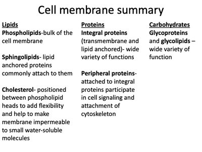
Cell Membrane Summary
The cell membrane is a complex, dynamic structure composed of lipids, proteins, and carbohydrates, each contributing to its selective permeability, signaling, and structural integrity.
Lipids: Phospholipids, sphingolipids, and cholesterol form the structural framework.
Proteins: Integral and peripheral proteins serve in transport, signaling, and structural roles.
Carbohydrates: Glycoproteins and glycolipids are involved in cell recognition and communication.
Additional info: The dynamic nature of the membrane allows cells to adapt to changing environments and maintain homeostasis.
