 Back
BackCell Structure and Function: Comprehensive Study Notes
Study Guide - Smart Notes
 Tailored notes based on your materials, expanded with key definitions, examples, and context.
Cell Structure and Function
Cell Theory
The cell theory is a fundamental concept in biology that outlines the properties and significance of cells in living organisms.
Cells are the building blocks of all plants and animals.
Cells are the smallest functioning units of life.
Cells are produced through the division of preexisting cells.
Each cell maintains homeostasis, regulating its internal environment.
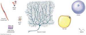
Diversity of Cells in the Human Body
Human bodies contain trillions of cells, each specialized for different functions and maintaining homeostasis through coordinated action. Cells vary greatly in shape and size, reflecting their diverse roles.
Examples: Neurons, muscle cells, blood cells, fat cells, bone cells, and epithelial cells.
The Study of Cells (Cytology)
Cytology is the study of cell structure and function, primarily using microscopy.
Light Microscopy (LM): Uses visible light to observe cells.
Electron Microscopy (EM): Uses electron beams for higher resolution.
Transmission Electron Microscopy (TEM): Views internal cell structures.
Scanning Electron Microscopy (SEM): Views cell surfaces in detail.
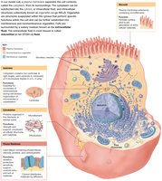
Cell Anatomy
Overview of Cell Anatomy
Despite their diversity, all cells share certain structural features, including a plasma membrane that separates the cytoplasm from the extracellular environment.
Plasma membrane: Boundary of the cell.
Cytoplasm: Internal fluid containing organelles.
Plasma Membrane Structure and Function
The plasma membrane is a dynamic structure that controls the movement of substances into and out of the cell.
Physical isolation from the extracellular fluid.
Regulation of exchange with the environment.
Sensitivity to environmental changes.
Structural support for the cell.
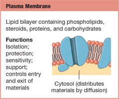
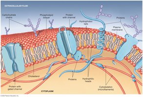
Components of the Plasma Membrane
Lipids: Phospholipids form a bilayer, with hydrophilic heads facing outward and hydrophobic tails inward. Cholesterol adds stiffness and reduces permeability.
Proteins: Integral (transmembrane) and peripheral proteins serve as channels, carriers, receptors, enzymes, anchors, and identifiers.
Carbohydrates: Attach to proteins (glycoproteins) or lipids (glycolipids) for cell recognition and adhesion.
Membrane Transport
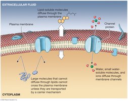
Permeability and Movement Across the Membrane
Membrane permeability determines which substances can cross. The plasma membrane is selectively permeable, allowing some substances to pass while restricting others.
Passive processes: Do not require energy (e.g., diffusion, osmosis, facilitated diffusion).
Active processes: Require energy, usually from ATP (e.g., active transport, vesicular transport).
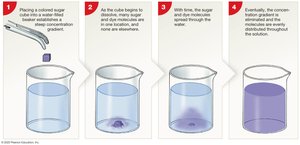
Diffusion
Diffusion is the movement of molecules from an area of high concentration to low concentration, driven by the concentration gradient.
Occurs until equilibrium is reached.
Can occur directly through the lipid bilayer or via channel proteins.
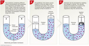
Osmosis
Osmosis is the diffusion of water across a selectively permeable membrane, from low solute concentration to high solute concentration.
Osmotic pressure: The force required to oppose water movement.
Water moves toward higher solute concentration.
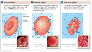
Tonicity
Tonicity describes the effect of solute concentration on cell shape:
Isotonic: No net water movement; cell retains normal shape.
Hypotonic: Water enters cell; cell swells and may burst (hemolysis in RBCs).
Hypertonic: Water leaves cell; cell shrivels (crenation in RBCs).
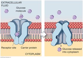
Carrier-Mediated Transport
Carrier proteins move specific substances across the membrane, either passively (facilitated diffusion) or actively (active transport).
Specificity: Each carrier transports certain molecules only.
Cotransport: Two substances move in the same direction.
Countertransport: Two substances move in opposite directions.
Facilitated Diffusion
Uses carrier proteins to move substances down their concentration gradient without energy input.
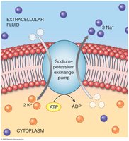
Active Transport
Requires ATP to move substances against their concentration gradient. The sodium-potassium exchange pump is a key example, maintaining cellular ion balance.
Ion pumps: Move ions like Na+, K+, Ca2+, Mg2+, Cl-.
Exchange pumps: Use ATP to move ions in opposite directions.
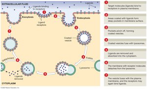
Vesicular Transport
Vesicular transport moves materials in and out of cells via membrane-bound vesicles. It includes endocytosis (into the cell) and exocytosis (out of the cell).
Endocytosis: Includes receptor-mediated endocytosis, pinocytosis, and phagocytosis.
Exocytosis: Releases substances from the cell.
Receptor-Mediated Endocytosis
Specific molecules bind to receptors, triggering vesicle formation and internalization.
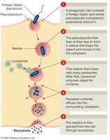
Phagocytosis
Specialized cells engulf large particles or pathogens, forming vesicles that fuse with lysosomes for digestion.
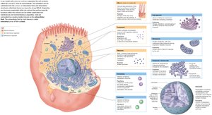
Cellular Components and Organelles
Cytoplasm and Cytosol
The cytoplasm is the material between the plasma membrane and the nucleus, containing cytosol (intracellular fluid) and organelles.
Cytosol: Contains dissolved nutrients, ions, proteins, and waste products.
Inclusions: Insoluble materials stored in the cytosol.
Organelles
Organelles are specialized structures that perform specific cellular functions. They are classified as membranous (isolated from cytosol) or nonmembranous (in direct contact with cytosol).
Cytoskeleton
The cytoskeleton provides structural support and facilitates movement within the cell.
Microfilaments: Thin filaments of actin, involved in cell movement and shape.
Intermediate filaments: Provide strength and stability.
Microtubules: Hollow tubes of tubulin, form spindle fibers and move organelles.
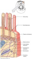
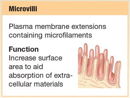
Microvilli
Microvilli are finger-like extensions of the plasma membrane that increase surface area for absorption.
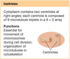
Centrioles
Centrioles are cylindrical structures involved in organizing microtubules during cell division.
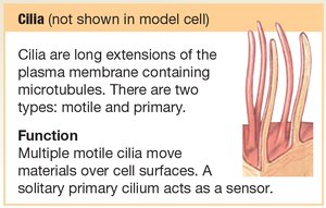
Cilia and Flagella
Cilia are long extensions that move substances across the cell surface; flagella move the cell itself.
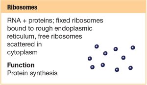
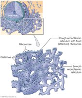
Ribosomes
Ribosomes are the sites of protein synthesis, found free in the cytoplasm or attached to the rough endoplasmic reticulum.
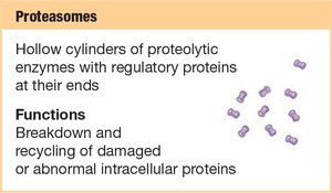
Proteasomes
Proteasomes are complexes that degrade and recycle damaged or abnormal proteins.
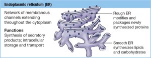
Endoplasmic Reticulum (ER)
The ER is a network of membranes involved in synthesis, storage, and transport.
Smooth ER (SER): Synthesizes lipids and carbohydrates.
Rough ER (RER): Studded with ribosomes; synthesizes and packages proteins.
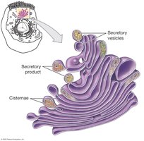
Golgi Apparatus
The Golgi apparatus modifies, packages, and distributes proteins and lipids.
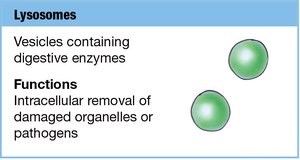
Lysosomes
Lysosomes are vesicles containing digestive enzymes for intracellular digestion and recycling.
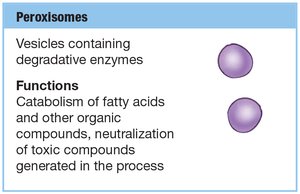
Peroxisomes
Peroxisomes contain enzymes that break down fatty acids and neutralize toxic compounds.
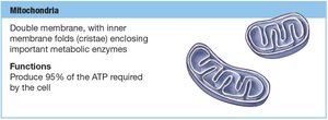
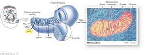
Mitochondria
Mitochondria are the powerhouses of the cell, generating ATP through aerobic metabolism.
Double membrane: Outer membrane and highly folded inner membrane (cristae).
Matrix: Site of metabolic reactions.
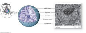
The Nucleus
Nuclear Structure and Function
The nucleus is the control center of the cell, containing DNA and directing cellular activities.
Nuclear envelope: Double membrane with nuclear pores for molecular exchange.
Nucleoli: Sites of ribosomal RNA synthesis.
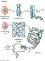
Chromatin: Loosely coiled DNA in non-dividing cells; condenses into chromosomes during division.
Genetic Information and Protein Synthesis
DNA contains the genetic code, which is transcribed and translated to produce proteins.
Gene: A segment of DNA coding for a specific protein.
Triplet code: Three DNA bases code for one amino acid.
Transcription
Transcription is the synthesis of messenger RNA (mRNA) from a DNA template.
RNA polymerase binds to the promoter region and synthesizes mRNA.
Uracil replaces thymine in RNA.
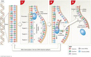
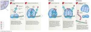
Translation
Translation is the process by which ribosomes use mRNA to assemble amino acids into proteins.
Initiation: Ribosome assembles at the start codon (AUG).
Elongation: tRNA brings amino acids, which are joined by peptide bonds.
Termination: Ribosome reaches a stop codon and releases the polypeptide.

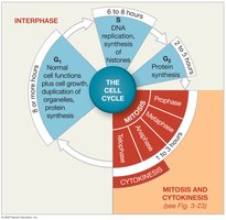
Cell Life Cycle
Stages of the Cell Life Cycle
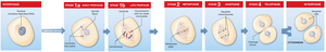
The cell life cycle includes interphase, mitosis, and cytokinesis, ensuring growth, repair, and reproduction.
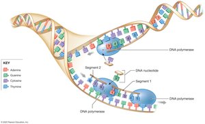
Interphase: Cell grows, duplicates organelles, and replicates DNA (G1, S, G2 phases).
Mitosis: Division of the nucleus (prophase, metaphase, anaphase, telophase).
Cytokinesis: Division of the cytoplasm, forming two daughter cells.
Cell Division and Cancer
Uncontrolled cell division can lead to tumor formation. Benign tumors remain localized, while malignant tumors invade tissues and metastasize. Cancer is characterized by gene mutations and abnormal cell growth.
Cellular Differentiation
Differentiation is the process by which cells become specialized, turning off certain genes to perform specific functions. This leads to the formation of tissues with distinct roles in the body.
