 Back
BackCell Structure and Function: Mini-Textbook Study Notes
Study Guide - Smart Notes
 Tailored notes based on your materials, expanded with key definitions, examples, and context.
Cell Structure and Function
Cell Basics
Cells are the smallest functional units of life, responsible for carrying out all chemical activities necessary to sustain living organisms. Their microscopic structure is closely related to their function, and they are organized into tissues and organs, forming the basis for higher-level functions in the body.
Cytology: The study of cells.
Cells carry out all chemical activities needed for life, including forming macromolecules and producing enzymes.
Clinical vital signs reflect cellular activity.
Despite their diversity, most cells share common features.
Cell Theory
The cell theory is a fundamental concept in biology, stating:
All living things are composed of cells.
All cells arise from pre-existing cells.
Cells are the basic units of life.
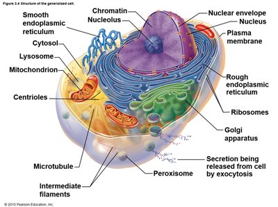
Generalized Cell Features
Most animal cells share three main features:
Nucleus: Contains genetic material and controls cellular activities.
Cytoplasm: The interior space filled with cytosol, a gel-like substance.
Plasma Membrane: A boundary that regulates interactions between the cell's interior and exterior.
Cell Membrane Structure
Plasma Membrane and the Fluid Mosaic Model
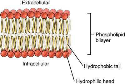
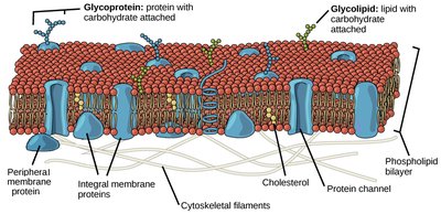
The plasma membrane is a selectively permeable barrier composed primarily of a phospholipid bilayer. It regulates the movement of substances into and out of the cell and maintains the membrane potential (charge difference across the membrane).
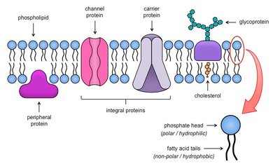
Phospholipid Bilayer: Hydrophilic heads face outward, hydrophobic tails face inward.
Fluid Mosaic Model: The membrane is flexible (fluid) and contains a mosaic of proteins and other molecules.
Cholesterol: Stabilizes the membrane and adjusts its flexibility.
Proteins: Integral (span the membrane) or peripheral (attached to the surface); function as receptors, channels, anchors, enzymes, and in cell identification.
Functions of Membrane Proteins
Transport: Channel and carrier proteins move substances across the membrane.
Receptors: Bind specific molecules (e.g., hormones) and trigger cellular responses.
Enzymatic Activity: Catalyze chemical reactions at the membrane surface.
Cell-Cell Recognition: Glycoproteins serve as identification tags.
Attachment: Anchor the membrane to the cytoskeleton and extracellular matrix.
Cell-to-Cell Joining: Form intercellular junctions for communication and adhesion.
Membrane Transport
Passive Transport
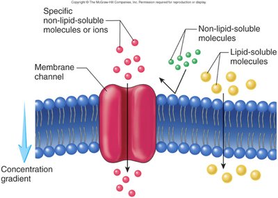
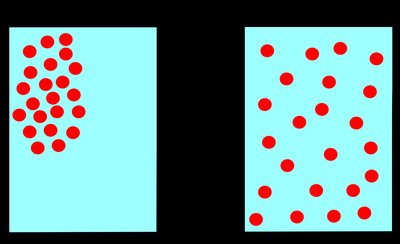
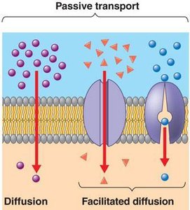
Passive transport involves the movement of substances across the membrane without the use of cellular energy (ATP). Substances move down their concentration gradient.
Simple Diffusion: Small, nonpolar molecules (e.g., O2, CO2) move directly through the lipid bilayer.
Facilitated Diffusion: Larger or polar molecules (e.g., glucose, ions) move through specific transport proteins.
Osmosis: Diffusion of water through aquaporins, following solute concentration (osmolarity).
Active Transport
Active transport requires energy (ATP) to move substances against their concentration gradient (from low to high concentration).
Primary Active Transport: Direct use of ATP to transport molecules (e.g., sodium-potassium pump).
Secondary Active Transport: Uses the energy from the movement of one substance down its gradient to move another substance up its gradient.
Vesicular Transport
Large molecules or particles are transported across the membrane in vesicles, a process that requires energy.
Endocytosis: Uptake of substances into the cell via vesicle formation.
Phagocytosis: "Cell eating"; engulfment of large particles.
Pinocytosis: "Cell drinking"; uptake of fluids.
Receptor-Mediated Endocytosis: Specific uptake of molecules via receptor binding.
Exocytosis: Expulsion of substances from the cell via vesicles.
Cytoplasm and Organelles
Cytoplasm
The cytoplasm is the region between the plasma membrane and the nucleus, containing cytosol (fluid) and organelles. It is the site of most cellular activities.
Cytosol: The fluid component, mostly water with dissolved solutes.
Inclusions: Non-living chemical substances (e.g., pigments, fat droplets).
Organelles
Organelles are specialized structures within the cell, each with specific functions essential for cell survival and activity.
Mitochondria: Powerhouse of the cell; site of ATP production via aerobic and anaerobic respiration.
Endoplasmic Reticulum (ER): Network of membranes; rough ER has ribosomes for protein synthesis, smooth ER is involved in lipid metabolism and detoxification.
Ribosomes: Sites of protein synthesis; can be free in cytoplasm or attached to rough ER.
Golgi Apparatus: Modifies, sorts, and packages proteins and lipids for secretion or delivery to other organelles.
Lysosomes: Contain digestive enzymes to break down macromolecules and cellular debris.
Peroxisomes: Detoxify harmful substances and neutralize free radicals.
Cytoskeleton: Network of protein filaments (microtubules, intermediate filaments, microfilaments) that provide structural support, cell shape, and movement.
Centrioles: Involved in cell division and organization of microtubules.
Cellular Extensions: Cilia (movement of substances), flagella (cell movement), microvilli (increase surface area).
Nucleus: The Control Center
The nucleus contains most of the cell's genetic material and directs all cellular activities.
Nuclear Envelope: Double membrane with pores for molecular exchange.
Nucleolus: Site of ribosome and rRNA synthesis.
Chromatin: DNA wrapped around histone proteins; condenses into chromosomes during cell division.
Cell Membrane Junctions
Most cells are bound together in tissues by specialized junctions that facilitate communication and adhesion.
Tight Junctions: Create a seal between adjacent cells to prevent leakage.
Gap Junctions: Allow direct communication between cells via connexons.
Desmosomes: Anchor cells together using protein plaques and intermediate filaments.
Osmosis and Tonicity
Osmosis is the passive diffusion of water across a selectively permeable membrane. Tonicity describes the relative concentration of solutes on either side of the membrane, affecting cell volume and function.
Isotonic: Equal solute concentration inside and outside the cell; no net water movement.
Hypotonic: Lower solute concentration outside the cell; water enters the cell, which may swell.
Hypertonic: Higher solute concentration outside the cell; water leaves the cell, which may shrink.
Clinical Connections
Hypercholesterolemia: LDLs cannot enter cells via endocytosis, leading to high blood cholesterol and arteriosclerosis.
Type 2 Diabetes: Reduced exocytosis of insulin impairs blood glucose regulation.
