 Back
Cell Structure, DNA, Chromosomes, and Cell Division: Study Notes for ANP College
Study Guide - Smart Notes
 Tailored notes based on your materials, expanded with key definitions, examples, and context.
The Nucleus and Its Components
Nuclear Envelope
The nuclear envelope is a double membrane structure that surrounds the nucleus in eukaryotic cells. It contains numerous nuclear pores that regulate the movement of molecules between the nucleus and cytoplasm.
Function: Separates nuclear contents from cytoplasm; controls entry and exit of proteins, RNA, and other molecules.
Nucleolus
The nucleolus is a dense region within the nucleus responsible for ribosome assembly.
Function: Synthesizes ribosomal RNA (rRNA) and assembles ribosome subunits.
Nucleoplasm
The nucleoplasm is the fluid matrix inside the nucleus, containing chromatin and the nucleolus.
Function: Supports nuclear structures and provides a medium for molecular transport.
Chromatin and Chromosomes
Chromatin is the complex of DNA and proteins found in the nucleus. During cell division, chromatin condenses to form chromosomes.
Chromatin: All DNA and associated proteins in the nucleus.
Chromosome: A distinct piece of chromatin; each chromosome contains genes coding for proteins.
Genes
A gene is a segment of DNA that provides instructions for synthesizing a specific protein. Genes contain promoter and terminator regions to signal the start and end of transcription.
Promoter: Start signal for transcription.
Terminator: Stop signal for transcription.

Human Chromosomes
Chromosome Structure and Number
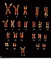
Humans have 46 chromosomes arranged in 23 pairs. Each pair consists of homologous chromosomes, one from each parent. Adult cells are diploid, containing two copies of each chromosome.
Homologous chromosomes: Chromosome pairs that carry genes for the same traits.
Diploid: Two sets of chromosomes (2n).
DNA Replication
Overview of DNA Replication
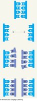
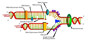
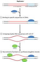
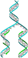
DNA replication is the process of copying the DNA molecule before cell division. It occurs during the S phase of interphase and follows a semiconservative model, where each new DNA molecule contains one original strand and one new strand.
Semiconservative replication: Each daughter DNA molecule retains half of the original DNA.
Key steps: DNA strands separate, new nucleotides pair with each template strand, forming two identical DNA molecules.
Cell Division
The Cell Cycle
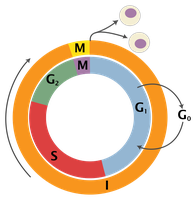
The cell cycle describes the sequence of events in cell growth and division. It consists of interphase (G1, S, G2) and the mitotic phase (M).
G1 phase: Cell growth and preparation for DNA replication.
S phase: DNA synthesis (replication).
G2 phase: Preparation for mitosis.
M phase: Mitosis and cytokinesis.
Mitosis
Mitosis is the process by which somatic cells divide, producing two genetically identical daughter cells. It is essential for growth, repair, and maintenance.
Phases: Prophase, Metaphase, Anaphase, Telophase.
Result: Two diploid cells.
Meiosis
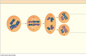
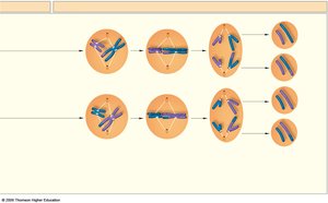
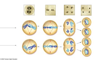
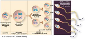
Meiosis is the cell division process that produces gametes (sperm and egg) with half the chromosome number (haploid). It involves two rounds of division: Meiosis I and Meiosis II.
Meiosis I: Separates homologous chromosome pairs.
Meiosis II: Separates sister chromatids.
Result: Four haploid cells, each genetically unique.
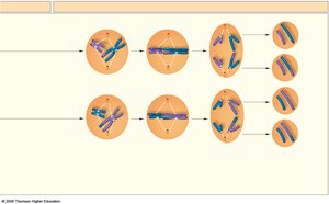
Stages of Meiosis
Prophase I: Chromosomes condense, crossing-over occurs.
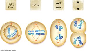
Metaphase I: Homologous pairs align at the cell midline.
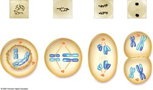
Anaphase I: Homologous chromosomes separate.
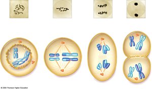
Telophase I: Cells divide, chromosomes remain X-shaped.
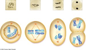
Prophase II: Chromosomes prepare for second division.
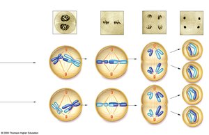
Metaphase II: Chromosomes align at midline.
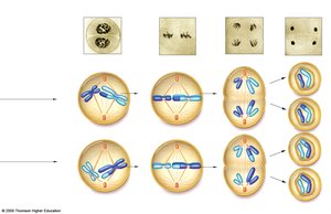
Anaphase II: Sister chromatids separate.
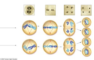
Telophase II: Four haploid gametes form.
Crossing Over
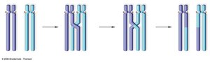
Crossing over is the exchange of genetic material between homologous chromosomes during Prophase I of meiosis. This increases genetic variation in offspring.
Gametes: Sperm and Egg
Sperm
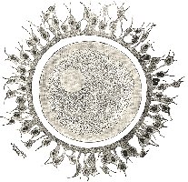
Sperm are motile male gametes, containing a nucleus, flagella, and mitochondria. They deliver DNA to the egg during fertilization.
Egg (Ovum)
The egg is the female gamete, much larger than sperm, and provides organelles and cytoplasm for the zygote. Only one of four cells produced in meiosis survives as the egg.
Apoptosis
Programmed Cell Death
Apoptosis is the process of programmed cell death, essential for development and tissue homeostasis. Cells undergoing apoptosis shrink, fragment, and are removed by phagocytes.
Transcription and Translation
Transcription
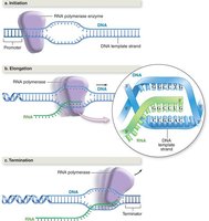
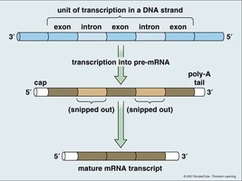
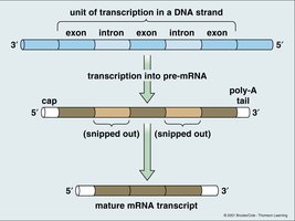
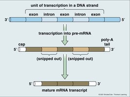
Transcription is the process of synthesizing RNA from a DNA template. The cell reads one strand of DNA and produces a complementary RNA strand, which is then modified to become mature mRNA.
Key steps: Initiation, elongation, termination.
Introns: Non-coding regions removed from pre-mRNA.
Exons: Coding regions retained in mature mRNA.
RNA Types
Ribosomal RNA (rRNA): Forms part of the ribosome, catalyzes protein synthesis.
Transfer RNA (tRNA): Transfers amino acids to the ribosome during translation.
Messenger RNA (mRNA): Carries genetic information from DNA to ribosome.
Translation
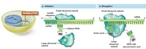
Translation is the process of synthesizing a protein from mRNA. Ribosomes read mRNA codons, tRNA brings corresponding amino acids, and the ribosome links them together.
Start codon: AUG (methionine).
Stop codons: UAA, UAG, UGA.
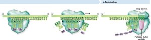
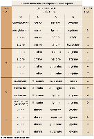
RNA Codon Chart
The codon chart is used to determine which amino acid corresponds to each three-nucleotide sequence (codon) in mRNA.
Summary Table: Cell Division Comparison
The following table compares mitosis and meiosis:
Feature | Mitosis | Meiosis |
|---|---|---|
Number of divisions | 1 | 2 |
Number of daughter cells | 2 | 4 |
Chromosome number | Diploid (2n) | Haploid (n) |
Genetic variation | None | High (crossing over, independent assortment) |
Function | Growth, repair | Gamete production |
Key Equations
Semiconservative DNA Replication
Each new DNA molecule consists of one old strand and one new strand:
Central Dogma of Molecular Biology
Describes the flow of genetic information:
Chromosome Number in Gametes
Gametes have half the chromosome number of somatic cells:
Additional info:
Some diagrams and images were included to visually reinforce key concepts such as chromosome structure, DNA replication, meiosis stages, and transcription/translation.
Definitions and explanations were expanded for clarity and completeness.
