 Back
Cells and Tissues: Structure, Classification, and Function
Study Guide - Smart Notes
 Tailored notes based on your materials, expanded with key definitions, examples, and context.
Introduction to Cells and Tissues
Overview of Human Body Tissues
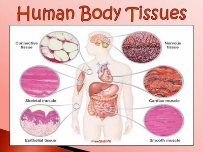
The human body is composed of over 75 trillion cells, which are organized into approximately 200 different types. These cells are grouped into four primary tissue types, each with distinct structures and functions: epithelial tissue, connective tissue, muscular tissue, and neural tissue. Understanding these tissues is foundational for the study of anatomy and physiology.
Levels of Organization in the Body
From Molecules to Organ Systems
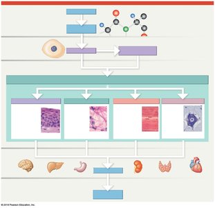
The body is organized hierarchically, starting from the chemical level (atoms and molecules), progressing to cells, tissues, organs, and finally organ systems. Each level builds upon the previous, with tissues forming the basic structural and functional units of organs.
Epithelial Tissue
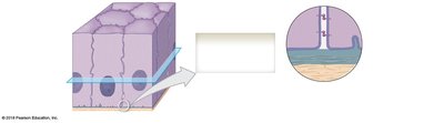
Characteristics of Epithelial Tissue
Cellularity: Epithelial cells are closely packed with minimal intercellular space.
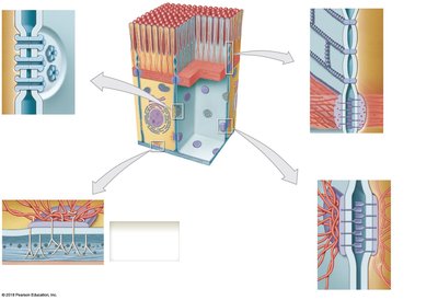
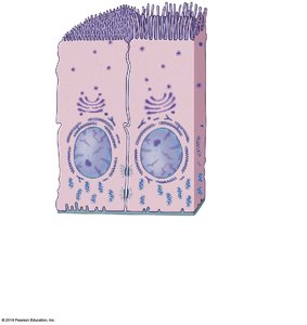
Polarity: Cells have an exposed apical surface and an attached basal surface, which differ structurally and functionally.
Attachment: The basal surface is attached to a basement membrane (basal lamina and reticular lamina).
Avascularity: Epithelial tissues lack blood vessels.
Regeneration: High capacity for renewal via stem cell division near the basal lamina.
Functions of Epithelial Tissue
Physical protection: Shields underlying tissues from abrasion, dehydration, and destruction.
Control of permeability: Regulates entry and exit of substances.
Provides sensation: Contains sensory nerve endings (neuroepithelia).
Produces secretions: Forms glands for secretion of substances.
Specializations of Epithelial Cells
Microvilli: Increase surface area for absorption (e.g., digestive tract).
Stereocilia: Long microvilli found in the inner ear and male reproductive tract.
Cilia: Move substances over the apical surface (e.g., respiratory tract).
Classification of Epithelia
By Layers:
Simple epithelium: One layer of cells; found in protected areas.
Stratified epithelium: Two or more layers; found in areas of mechanical or chemical stress.
By Cell Shape:
Squamous: Flat cells.
Cuboidal: Cube-shaped cells.
Columnar: Taller than wide, column-shaped cells.
Examples of Epithelial Types
Simple Squamous Epithelium: Lines body cavities, heart, blood vessels; reduces friction, absorption, secretion.
Stratified Squamous Epithelium: Surface of skin, mouth, esophagus; protection against abrasion and pathogens.
Simple Cuboidal Epithelium: Kidney tubules, glands; secretion and absorption.
Stratified Cuboidal Epithelium: Rare, found in sweat gland ducts; protection, secretion, absorption.
Simple Columnar Epithelium: Lining of stomach, intestines; protection, secretion, absorption.
Stratified Columnar Epithelium: Pharynx, anus, mammary glands; protection.
Pseudostratified Ciliated Columnar Epithelium: Respiratory tract; protection, secretion.
Transitional Epithelium: Urinary bladder; permits expansion and recoil.
Glandular Epithelia
Exocrine Glands: Secrete via ducts to epithelial surface (serous, mucous, or mixed secretions).
Endocrine Glands: Secrete hormones into blood or lymph.
Unicellular Glands: Goblet and mucous cells (e.g., in trachea, salivary glands).
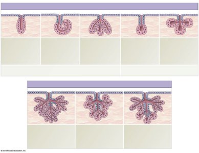
Multicellular Glands: Secretory sheets (e.g., stomach lining).
Structural Types: Tubular, alveolar (acinar), tubuloalveolar; simple (no branching) or compound (branched).
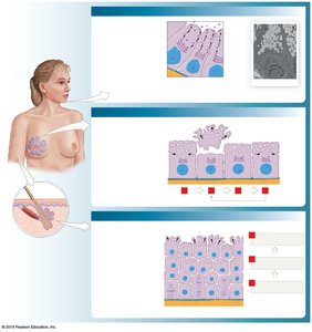
Modes of Secretion:
Eccrine (Merocrine): Exocytosis (e.g., salivary glands).
Apocrine: Shedding of apical cytoplasm (e.g., mammary glands).
Holocrine: Cell bursts, releasing contents (e.g., sebaceous glands).
Connective Tissue
General Features and Functions
Connective tissues are characterized by specialized cells, extracellular protein fibers, and a ground substance (matrix). The matrix can be fluid, gel-like, or solid, depending on the tissue type. Functions include structural support, transport, protection, energy storage, and defense against pathogens.
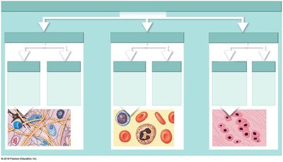
Classification of Connective Tissues
Connective Tissue Proper: Loose (areolar, adipose, reticular) and dense (regular, irregular, elastic).
Fluid Connective Tissue: Blood and lymph.
Supporting Connective Tissue: Cartilage (hyaline, elastic, fibrous) and bone.
Cells of Connective Tissue Proper
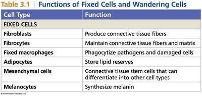
Cell Type | Function |
|---|---|
Fibroblasts | Produce connective tissue fibers |
Fibrocytes | Maintain connective tissue fibers and matrix |
Fixed macrophages | Phagocytize pathogens and damaged cells |
Adipocytes | Store lipid reserves |
Mesenchymal cells | Stem cells that differentiate into other cell types |
Melanocytes | Synthesize melanin |
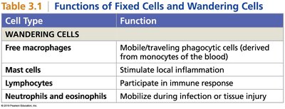
Free macrophages | Mobile phagocytic cells |
Mast cells | Stimulate local inflammation |
Lymphocytes | Participate in immune response |
Neutrophils and eosinophils | Mobilize during infection or injury |
Fibers of Connective Tissue Proper
Collagen fibers: Provide tensile strength.
Reticular fibers: Form supportive networks.
Elastic fibers: Contain elastin, allowing stretch and recoil.
Loose Connective Tissues
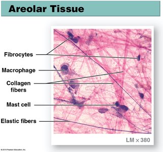
Areolar Tissue: Connects skin to muscle; provides minimal support and independent movement.
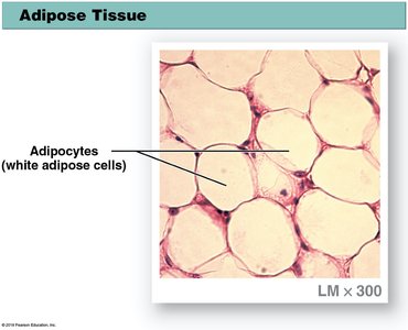
Adipose Tissue: Cushions, insulates, and stores energy; found in hypodermis, buttocks, around organs.
Reticular Tissue: Forms supporting framework in liver, spleen, lymph nodes, and bone marrow.
Dense Connective Tissues
Dense Regular: Tendons (muscle to bone), ligaments (bone to bone), aponeuroses (muscle to muscle).
Dense Irregular: Forms sheaths around nerves and muscles, fibrous capsules around organs.
Elastic Tissue: Stabilizes vertebrae, allows stretching.
Fluid Connective Tissues
Blood: Contains erythrocytes (O2 and CO2 transport), leukocytes (immune defense), and platelets (clotting); matrix is plasma.
Lymph: Similar to blood plasma, involved in immune response.
Supporting Connective Tissues
Cartilage: Chondrocytes in lacunae, avascular, surrounded by perichondrium; types include hyaline (joints, trachea), elastic (ear, epiglottis), and fibrous (intervertebral discs, pubic symphysis).
Bone: Osteocytes in lacunae, solid matrix (lamellae), covered by periosteum; types include compact and spongy bone.
Membranes
Types of Membranes
Mucous Membranes: Line digestive, respiratory, reproductive, and urinary tracts; resist pathogen entry, keep surfaces moist.
Serous Membranes: Line body cavities; consist of parietal and visceral layers (pleura, peritoneum, pericardium).
Cutaneous Membrane: Skin; thick, waterproof, keratinized stratified squamous epithelium.
Synovial Membrane: Lines joint cavities; produces synovial fluid, lacks basal lamina, has gaps between cells.
Muscle Tissue
Types of Muscle Tissue
Skeletal Muscle: Multinucleated, striated, voluntary; moves skeleton, incapable of cell reproduction but can repair via myosatellite cells.
Cardiac Muscle: Single nucleus, striated, involuntary; found only in heart, connected by intercalated discs, relies on pacemaker cells.
Smooth Muscle: Single nucleus, nonstriated, involuntary; found in walls of blood vessels, digestive, respiratory, urinary, and reproductive tracts; capable of cell reproduction.
Nervous Tissue
Structure and Function
Neurons: Specialized for conduction of electrical impulses; consist of soma (cell body), axon, and dendrites; incapable of cell reproduction.
Neuroglia: Supporting cells; protect and support neurons.
Tissues and Aging
Effects of Aging on Tissues
Repair and maintenance become less efficient with age.
Epithelia thin, connective tissues become fragile.
Cardiac muscle and neural tissue cannot regenerate, leading to cumulative damage.
Bone strength decreases (osteoporosis).
