 Back
Cells: The Living Units – Cytoplasm, Organelles, and Nucleus
Study Guide - Smart Notes
 Tailored notes based on your materials, expanded with key definitions, examples, and context.
Cells: The Living Units
Cytoplasm
The cytoplasm is the cellular material located between the plasma membrane and the nucleus. It serves as the site for most cellular activities and contains three main components: cytosol, inclusions, and organelles.
Cytosol: Gel-like solution composed mainly of water and soluble molecules such as proteins, salts, and sugars.
Inclusions: Insoluble molecules that vary with cell type (e.g., glycogen granules, pigments, lipid droplets, vacuoles, crystals).
Organelles: Specialized structures within the cell, each with distinct functions. Organelles are classified as either membranous or nonmembranous.
Cytoplasmic Organelles
Organelles are the metabolic machinery of the cell, enabling compartmentalization and specialized functions. They are divided into two categories:
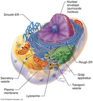
Membranous Organelles: Mitochondria, endoplasmic reticulum, Golgi apparatus, peroxisomes, lysosomes.
Nonmembranous Organelles: Ribosomes, cytoskeleton, centrioles.
Mitochondria
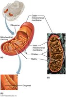
Mitochondria are known as the "power plants" of the cell, producing most of the cell's ATP via aerobic respiration. They are enclosed by double membranes, with the inner membrane forming folds called cristae, which are embedded with proteins essential for cellular respiration. Mitochondria contain their own DNA, RNA, and ribosomes, and can divide by fission.
Ribosomes
Ribosomes are nonmembranous organelles responsible for protein synthesis. They consist of protein and ribosomal RNA (rRNA) and exist in two forms:
Free ribosomes: Float freely in the cytosol and synthesize soluble proteins for use within the cell.
Membrane-bound ribosomes: Attached to the endoplasmic reticulum, synthesizing proteins for membranes, lysosomes, or export.
Endoplasmic Reticulum (ER)
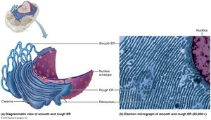
The ER is a network of interconnected membranous tubes continuous with the nuclear envelope. It is divided into two types:
Rough ER: Studded with ribosomes; site of synthesis and modification of proteins destined for secretion, membranes, or lysosomes.
Smooth ER: Lacks ribosomes; involved in lipid metabolism, detoxification, glycogen breakdown, and calcium storage.
Golgi Apparatus
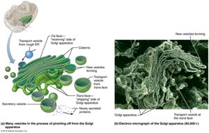
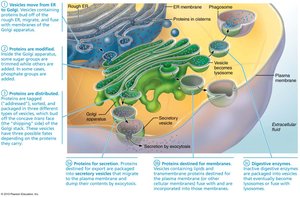
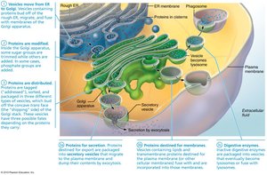
The Golgi apparatus consists of stacked, flattened membranous sacs. It modifies, concentrates, and packages proteins and lipids received from the rough ER. The Golgi directs products to one of three pathways: secretion, membrane insertion, or lysosomal storage.
Protein Processing and Distribution
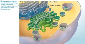
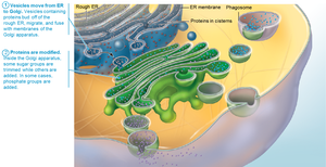
Proteins synthesized on the rough ER are processed and distributed through a sequence of events involving the ER, Golgi apparatus, and vesicles. Proteins are modified, sorted, and packaged for their final destinations.
Peroxisomes
Peroxisomes are membranous sacs containing enzymes that detoxify harmful substances. They neutralize free radicals by converting them to hydrogen peroxide (H2O2), which is then converted to water by catalase. Peroxisomes also participate in fatty acid metabolism.
Lysosomes
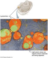
Lysosomes are spherical membranous bags containing digestive enzymes. They digest bacteria, viruses, toxins, and nonfunctional organelles, and play roles in metabolic functions such as glycogen breakdown and calcium release. Lysosomal storage diseases, such as Tay-Sachs, result from defective lysosomal enzymes.
Endomembrane System
The endomembrane system includes the ER, Golgi apparatus, vesicles, lysosomes, nuclear envelope, and plasma membrane. These structures work together to produce, degrade, store, and export biological molecules, and to degrade harmful substances.
Cytoskeleton
The cytoskeleton is an elaborate network of protein rods that provide structural support, facilitate movement, and organize cell components. It consists of three types:
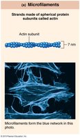
Microfilaments: Thinnest, composed of actin; involved in cell shape, motility, and membrane activities.
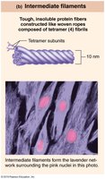
Intermediate filaments: Tough, ropelike fibers; provide tensile strength and resist pulling forces.
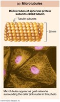
Microtubules: Largest, hollow tubes of tubulin; determine cell shape, organelle distribution, and serve as tracks for motor proteins.
Centrosome and Centrioles
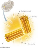
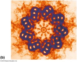
The centrosome is a microtubule organizing center near the nucleus, containing centrioles. Centrioles are barrel-shaped organelles that form the basis of cilia and flagella and are essential for cell division.
Cellular Extensions
Cilia and Flagella
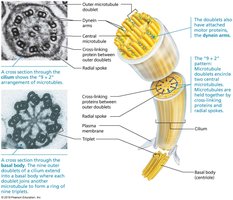
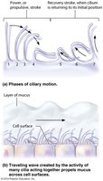
Cilia are motile extensions that move substances across cell surfaces, while flagella propel entire cells. Both are composed of microtubules arranged in a "9 + 2" pattern, synthesized by centrioles (basal bodies).
Microvilli
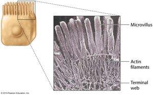
Microvilli are minute, fingerlike projections that increase surface area for absorption. They contain a core of actin microfilaments for structural support.
Nucleus
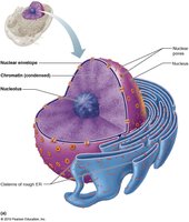
The nucleus is the largest organelle, containing the genetic library for protein synthesis. It responds to cellular signals and regulates gene expression. Most cells are uninucleate, but some are multinucleate or anucleate.
Structure of the Nucleus
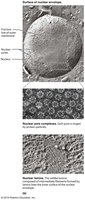
Nuclear envelope: Double-membrane barrier enclosing nucleoplasm; outer layer continuous with rough ER and studded with ribosomes; inner layer (nuclear lamina) maintains shape.
Nucleoli: Spherical bodies involved in rRNA synthesis and ribosome assembly.
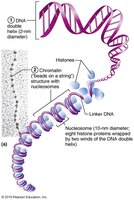
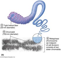
Chromatin: Composed of DNA, histone proteins, and RNA; organized into nucleosomes. Chromosomes are condensed chromatin, protecting DNA during cell division.
Summary Table: Major Cytoplasmic Organelles
Organelle | Structure | Main Function |
|---|---|---|
Mitochondria | Double membrane, cristae, own DNA | ATP production |
Ribosomes | Protein + rRNA, free or bound | Protein synthesis |
ER (Rough/Smooth) | Membranous tubes, rough with ribosomes | Protein/lipid synthesis, detoxification |
Golgi Apparatus | Stacked cisterns | Modification, packaging, sorting |
Peroxisomes | Membranous sacs | Detoxification, fatty acid metabolism |
Lysosomes | Membranous bags | Digestion, autolysis |
Cytoskeleton | Microfilaments, intermediate filaments, microtubules | Support, movement |
Centrioles | Barrel-shaped, microtubules | Cell division, cilia/flagella formation |
Nucleus | Envelope, nucleoli, chromatin | Genetic control, protein synthesis |
Additional info: The notes above expand on brief points with academic context, definitions, and examples to ensure completeness and clarity for college-level study.
