 Back
Cells: The Living Units – Plasma Membrane Structure & Function
Study Guide - Smart Notes
 Tailored notes based on your materials, expanded with key definitions, examples, and context.
Chapter 3: Cells – The Living Units
Introduction to Cells
Cells are the fundamental units of life, and their structure determines their function within multicellular organisms. The coordinated activity of all body cells is essential for the survival of the organism. According to the cell theory, all living things are composed of cells, and all cells arise from pre-existing cells through cell division (mitosis).
Cell Structure: Determines the specific function of each cell type.
Cell Division: Occurs via mitosis, ensuring continuity of life.
Multicellularity: Depends on the coordinated activity of specialized cells.
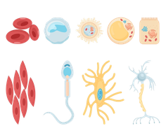
Example: Red blood cells transport oxygen, nerve cells transmit signals, and muscle cells contract to produce movement.
Generalized Cell Structure
Most animal cells share three main structural components:
Plasma Membrane: The outer boundary that separates the cell from its environment.
Cytoplasm: The region between the plasma membrane and nucleus, containing organelles and cytosol (intracellular fluid).
Nucleus: The control center containing genetic material (DNA).
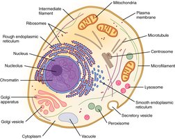
Additional info: Organelles such as mitochondria, endoplasmic reticulum, and Golgi apparatus perform specialized cellular functions.
Terminology: Prefixes for Cellular Location
Intra-: Within (e.g., intracellular – inside the cell)
Extra-: Outside (e.g., extracellular – outside the cell)
Inter-: Between (e.g., intercellular – between cells)
Extracellular Environment
Extracellular Matrix (ECM) and Fluids
The extracellular environment consists of the extracellular matrix (ECM) and various fluids that surround cells.
Extracellular Matrix (ECM): Non-fluid materials and interstitial fluid found between cells; provides structural and biochemical support.
Extracellular Fluid (ECF): All body fluid outside cells, including interstitial fluid, cerebrospinal fluid (CSF), serous fluid, synovial fluid, and blood plasma.
Plasma Membrane Structure
Lipid Bilayer
The plasma membrane is primarily composed of a lipid bilayer, which forms a selective barrier between the intracellular and extracellular environments.
Phospholipids: Main component; each molecule has a hydrophilic (water-loving) polar head and two hydrophobic (water-fearing) nonpolar fatty acid tails.
Arrangement: Hydrophilic heads face outward toward aqueous environments; hydrophobic tails face inward, away from water.
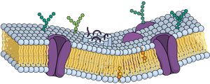
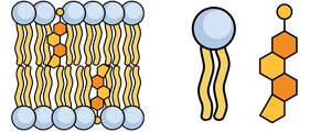
Additional info: Cholesterol molecules are interspersed within the bilayer, stabilizing membrane fluidity.
Membrane Proteins
Proteins embedded in or associated with the plasma membrane perform a variety of essential functions.
Integral Proteins: Firmly embedded; include transmembrane proteins that span the entire membrane.
Peripheral Proteins: Loosely attached to the membrane surface.
Functions of Membrane Proteins:
Junctions: Join cells together (e.g., tight junctions, desmosomes, gap junctions).
Enzymes: Catalyze chemical reactions.
Transport: Move substances across the membrane (channels, carriers).
Recognition: Serve as markers for cellular identification (glycoproteins).
Anchoring: Attach to cytoskeletal or extracellular structures.
Transduction: Receive and transmit signals into the cell.
Membrane Carbohydrates and the Glycocalyx
Carbohydrates attached to lipids (glycolipids) or proteins (glycoproteins) form the glycocalyx, a carbohydrate-rich area on the cell surface.
Function: Provides markers for cell-cell recognition and communication.
Fluid Mosaic Model
The plasma membrane is described by the fluid mosaic model:
Fluid: Components (phospholipids, proteins) are in constant motion.
Mosaic: The membrane is a patchwork of different molecules (lipids, proteins, carbohydrates).
Cell Junctions
Types of Cell Junctions
Specialized junctions connect adjacent cells and facilitate communication, adhesion, and barrier formation.
Tight Junctions: Impermeable; interlocking proteins create a watertight seal (e.g., between cells lining the digestive tract).
Desmosomes: Anchoring junctions; protein plaques and linker proteins provide mechanical strength (e.g., between skin cells).
Gap Junctions: Communicating junctions; protein channels (connexons) allow small molecules and ions to pass directly between cells (e.g., in cardiac and smooth muscle).
Plasma Membrane Transport
Passive Transport
Passive transport processes move substances across the membrane without energy input from the cell.
Filtration: Movement of water and solutes by hydrostatic pressure (e.g., across capillary walls).
Diffusion: Movement of molecules from high to low concentration (down their concentration gradient).
Simple Diffusion: Direct passage of nonpolar, fat-soluble molecules (e.g., O2, CO2).
Facilitated Diffusion: Passage of polar or charged molecules via protein channels or carriers (e.g., glucose, ions).
Osmosis: Diffusion of water across a selectively permeable membrane toward higher solute concentration.
Tonicity: The ability of a solution to change cell volume by osmosis.
Isotonic: Equal solute concentration; no net water movement.
Hypotonic: Lower solute concentration outside; water enters cell, causing swelling.
Hypertonic: Higher solute concentration outside; water leaves cell, causing shrinkage.
Active Transport
Active transport processes require cellular energy (usually ATP) to move substances against their concentration gradients.
Primary Active Transport: Direct use of ATP to transport substances (e.g., Na+/K+ pump).
Secondary Active Transport (Cotransport): Uses energy stored in concentration gradients created by primary active transport (e.g., Na+/glucose symport).
Vesicular Transport: Movement of large particles via vesicles (exocytosis and endocytosis).
Types of Endocytosis:
Phagocytosis: "Cell eating"; engulfment of solids.
Pinocytosis: "Cell drinking"; uptake of extracellular fluid.
Receptor-Mediated Endocytosis: Specific uptake of molecules via receptor proteins.
Membrane Potential and Cell Signaling
Membrane Potential
The membrane potential is the voltage difference across the plasma membrane, resulting from the unequal distribution of ions (mainly Na+ and K+).
Resting Membrane Potential (RMP): The steady-state voltage, typically negative inside the cell, maintained by the Na+/K+ pump and selective permeability of the membrane.
Plasma Membrane Receptors and Cell Signaling
Cells communicate via membrane receptors that respond to various signals:
Contact Signaling: Direct cell-to-cell recognition via membrane receptors; important in tissue development and immunity.
Electrical Signaling: Voltage-gated channels respond to changes in membrane potential; crucial in nerve and muscle cells.
Chemical Signaling: Ligand binding (e.g., neurotransmitters, hormones) activates membrane proteins, leading to intracellular signaling cascades.
G Protein-Coupled Receptors (GPCRs): A major class of membrane receptors that, upon ligand binding, activate G proteins to relay signals to effector proteins and generate second messengers, resulting in diverse cellular responses.
Summary Table: Types of Membrane Transport
Transport Type | Energy Required? | Direction | Examples |
|---|---|---|---|
Simple Diffusion | No | Down gradient | O2, CO2 |
Facilitated Diffusion | No | Down gradient | Glucose, ions |
Osmosis | No | Down water gradient | Water |
Primary Active Transport | Yes (ATP) | Against gradient | Na+/K+ pump |
Secondary Active Transport | Indirect (gradient) | Against gradient | Na+/glucose cotransport |
Vesicular Transport | Yes (ATP) | Bulk movement | Endocytosis, exocytosis |
