 Back
Cells: The Living Units – Structure and Function of the Plasma Membrane
Study Guide - Smart Notes
 Tailored notes based on your materials, expanded with key definitions, examples, and context.
Cells: The Living Units
Introduction to Cell Theory
Cells are the fundamental structural and functional units of all living organisms. The activities of an organism depend on the individual and collective activities of its cells. The structure of each cell is closely related to its function, and all cells arise from preexisting cells, ensuring the continuity of life.
Cell Theory: All living things are composed of cells; cells are the basic units of structure and function in living things; new cells are produced from existing cells.
Structure-Function Relationship: The shape and internal structures of a cell determine its biochemical activities.
Cell Diversity: The human body contains over 250 different types of cells, varying in size, shape, and function.
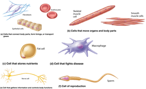
Generalized Cell Structure
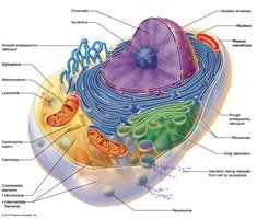
Despite their diversity, all human cells share three basic components:
Plasma Membrane: The flexible outer boundary that separates the cell from its environment.
Cytoplasm: The intracellular fluid containing organelles.
Nucleus: The control center containing DNA.
Extracellular Materials
Types of Extracellular Materials
Substances found outside cells are essential for cellular function and include:
Extracellular fluids: Interstitial fluid, blood plasma, and cerebrospinal fluid.
Cellular secretions: Such as saliva and mucus.
Extracellular matrix: A network of proteins and carbohydrates that provides structural support and acts as a "glue" to hold cells together.
Plasma Membrane Structure and Function
Overview of the Plasma Membrane
The plasma membrane acts as a dynamic barrier, separating the intracellular fluid (ICF) from the extracellular fluid (ECF). It controls what enters and exits the cell, maintaining homeostasis.
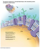
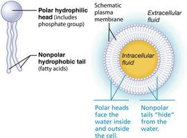
Fluid Mosaic Model: The membrane is composed of a phospholipid bilayer with embedded proteins, cholesterol, and carbohydrates, creating a constantly changing mosaic.
Membrane Lipids
The lipid bilayer forms the basic structure of the plasma membrane:
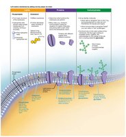
Phospholipids (75%): Have hydrophilic (water-loving) heads and hydrophobic (water-fearing) tails, forming a bilayer.
Glycolipids (5%): Lipids with attached sugar groups, found on the outer surface.
Cholesterol (20%): Stabilizes membrane fluidity and structure.
Membrane Proteins
Membrane proteins are crucial for communication and transport across the plasma membrane. They account for about half the mass of the membrane and are classified as:
Integral proteins: Firmly embedded in the membrane, often spanning the bilayer (transmembrane). Functions include transport, acting as enzymes, or receptors.
Peripheral proteins: Loosely attached to integral proteins, functioning as enzymes, motor proteins, or in cell-to-cell connections.
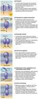
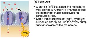
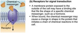
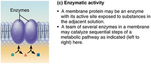
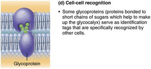
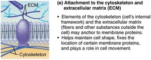
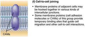
Table: Major Functions of Membrane Proteins
Function | Description |
|---|---|
Transport | Move substances across membrane (channels, carriers) |
Receptors for signal transduction | Bind chemical messengers and initiate cellular responses |
Enzymatic activity | Catalyze metabolic reactions |
Cell-cell recognition | Serve as identification tags for cell recognition |
Attachment to cytoskeleton/ECM | Maintain cell shape and stabilize membrane |
Cell-to-cell joining | Form intercellular junctions |
Glycocalyx
The glycocalyx is a carbohydrate-rich area on the cell surface, formed by glycoproteins and glycolipids. It serves as a biological marker for cell recognition and helps the immune system distinguish self from non-self.
Cell Junctions
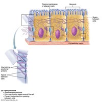
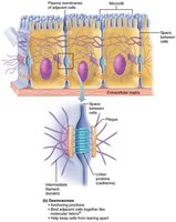
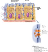
Most cells are bound together to form tissues and organs. The main types of cell junctions are:
Tight junctions: Form impermeable barriers to prevent leakage between cells.
Desmosomes: Provide anchoring strength, allowing cells to resist mechanical stress.
Gap junctions: Allow communication and passage of ions and small molecules between cells.
Membrane Transport
Selective Permeability
The plasma membrane is selectively permeable, allowing only certain molecules to cross. Substances move across the membrane by:
Passive transport: Does not require energy (ATP).
Active transport: Requires energy input (ATP).
Passive Membrane Transport
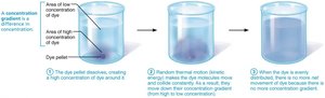
Passive transport involves the movement of substances down their concentration gradients. The main types are:
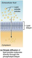
Simple diffusion: Movement of nonpolar, lipid-soluble substances directly through the lipid bilayer.
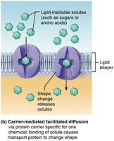
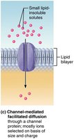
Facilitated diffusion: Movement of larger or polar molecules via protein carriers or channels.
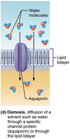
Osmosis: Diffusion of water across a selectively permeable membrane.
Factors Affecting Diffusion Speed
Concentration gradient: Greater differences increase diffusion rate.
Molecular size: Smaller molecules diffuse faster.
Temperature: Higher temperatures increase kinetic energy and diffusion rate.
Simple Diffusion
Nonpolar, lipid-soluble molecules (e.g., oxygen, carbon dioxide, steroid hormones) diffuse directly through the phospholipid bilayer.
Facilitated Diffusion
Certain hydrophilic molecules (e.g., glucose, amino acids, ions) are transported passively via:
Carrier-mediated: Substances bind to protein carriers, which change shape to move the molecule across.
Channel-mediated: Substances move through water-filled protein channels (e.g., aquaporins for water).
Osmosis
Osmosis is the movement of water across a selectively permeable membrane. Water moves from areas of low solute concentration (high water) to high solute concentration (low water) until equilibrium is reached.
Osmolarity and Tonicity
Osmolarity measures the total concentration of solute particles in a solution. Tonicity describes how a solution affects cell volume:
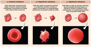
Isotonic: Same osmolarity as the cell; no net water movement.
Hypertonic: Higher osmolarity than the cell; water leaves the cell, causing it to shrink (crenation).
Hypotonic: Lower osmolarity than the cell; water enters the cell, causing it to swell and possibly burst (lysis).
Table: Effects of Tonicity on Red Blood Cells
Solution Type | Osmolarity | Effect on Cell |
|---|---|---|
Isotonic | Equal to cell | No change |
Hypertonic | Higher than cell | Cell shrinks (crenation) |
Hypotonic | Lower than cell | Cell swells/bursts (lysis) |
Diffusion and Osmosis Across Membranes
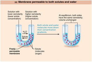
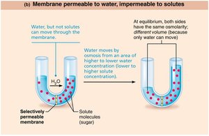
When solutions of different osmolarities are separated by a membrane:
If the membrane is permeable to both solutes and water, both diffuse until equilibrium is reached.
If the membrane is permeable only to water, only osmosis occurs, resulting in volume changes.
Osmotic and Hydrostatic Pressure
Water movement across membranes generates pressures:
Hydrostatic pressure: Outward pressure due to increased cell volume.
Osmotic pressure: Inward pressure due to water being drawn into areas of higher solute concentration.
In animal cells, excessive water entry can cause bursting, while water loss can cause shrinking.
Osmolarity Calculation
Osmolarity is calculated as:
For example, 1 M NaCl dissociates into Na+ and Cl−, so it is a 2 Osm solution.
