 Back
BackCells: The Living Units – Structure, Function, and Processes
Study Guide - Smart Notes
 Tailored notes based on your materials, expanded with key definitions, examples, and context.
Cell Theory and Overview
Introduction to Cells
Cells are the fundamental structural and functional units of all living organisms. The activities of an organism depend on the individual and collective activities of its cells. The structure of each cell determines its specific biochemical activities, and the continuity of life is maintained through cellular reproduction.
Cell Theory: All living things are composed of cells; cells are the basic units of structure and function; all cells arise from pre-existing cells.
Cell Diversity: Cells vary greatly in size, shape, and function, reflecting their specialized roles in the body.
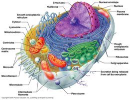
Structure of a Generalized Cell
Major Components
A typical eukaryotic cell consists of three main parts: the plasma membrane, cytoplasm (with organelles), and nucleus. Each component plays a vital role in maintaining cellular function and homeostasis.
Plasma Membrane: Separates the cell's internal environment from the external environment and regulates the movement of substances in and out of the cell.
Cytoplasm: Contains cytosol (fluid) and organelles, which perform various metabolic functions.
Nucleus: The control center of the cell, containing genetic material (DNA).
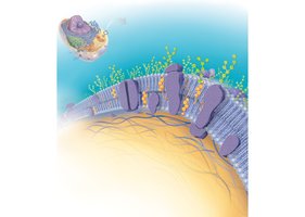
Plasma Membrane
Structure and Function
The plasma membrane is a dynamic structure composed of a double layer of phospholipids with embedded proteins, cholesterol, and glycolipids. It acts as a selective barrier and is involved in cell signaling, adhesion, and recognition.
Fluid Mosaic Model: Describes the flexible arrangement of lipids and proteins in the membrane.
Glycocalyx: A glycoprotein-rich area on the cell surface that serves as a biological marker for cell recognition.
Membrane Proteins: Integral and peripheral proteins perform functions such as transport, signaling, and cell adhesion.
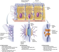
Membrane Junctions
Cells are connected by specialized junctions that maintain tissue integrity and facilitate communication.
Tight Junctions: Form impermeable barriers between cells (e.g., in the GI tract).
Desmosomes: Provide mechanical stability by anchoring cells together (e.g., in skin and heart muscle).
Gap Junctions: Allow direct communication between cells by permitting the passage of ions and small molecules (e.g., in heart muscle).
Membrane Transport
Selective Permeability
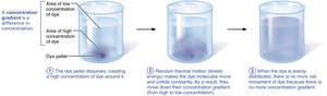
The plasma membrane is selectively permeable, allowing some substances to pass while restricting others. Transport mechanisms are classified as passive or active based on energy requirements.
Passive Processes: Do not require cellular energy (ATP); substances move down their concentration gradient.
Active Processes: Require ATP to move substances against their concentration gradient or via vesicular transport.
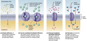
Passive Processes
Passive transport includes simple diffusion, facilitated diffusion, and osmosis.
Simple Diffusion: Movement of molecules from high to low concentration without assistance.
Facilitated Diffusion: Movement of molecules via protein channels or carriers.
Osmosis: Diffusion of water across a selectively permeable membrane, driven by solute concentration differences.
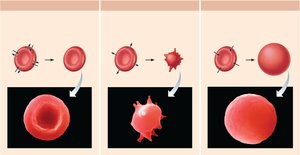
Osmosis and Tonicity
Osmosis is crucial for maintaining cell volume and function. Tonicity describes the effect of extracellular solutions on cell volume.
Isotonic Solution: No net movement of water; cell retains normal shape.
Hypertonic Solution: Water leaves the cell; cell shrinks (crenates).
Hypotonic Solution: Water enters the cell; cell swells and may burst (lyse).
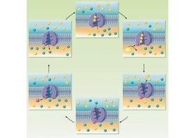
Active Processes
Active Transport
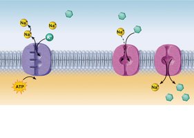
Active transport uses carrier proteins and ATP to move substances against their concentration gradients. Two main types are primary and secondary active transport.
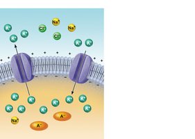
Primary Active Transport: Direct use of ATP to transport molecules (e.g., sodium-potassium pump).
Secondary Active Transport: Uses energy stored in ionic gradients created by primary active transport (e.g., cotransport of glucose with sodium).
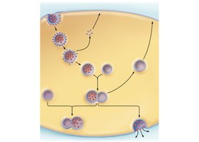
Vesicular Transport
Vesicular transport moves large particles and macromolecules across membranes using vesicles. This process requires energy (ATP).
Exocytosis: Transport of substances out of the cell (e.g., hormone secretion).
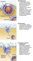
Endocytosis: Transport of substances into the cell (e.g., phagocytosis, pinocytosis, receptor-mediated endocytosis).
Transcytosis: Movement into, across, and out of the cell.
Vesicular Trafficking: Movement of substances within the cell.
Membrane Potential
Resting Membrane Potential (RMP)
The separation of oppositely charged ions across the plasma membrane creates a membrane potential, measured in millivolts (mV). The RMP is typically between –50 and –100 mV and is essential for nerve and muscle function.
Established by: Diffusion and active transport of ions, mainly potassium (K+).
Maintained by: Sodium-potassium pump and selective permeability of the membrane.
Cytoplasm and Organelles
Cytoplasm
The cytoplasm is the cellular material between the plasma membrane and the nucleus. It consists of cytosol (fluid), organelles (metabolic machinery), and inclusions (stored nutrients or pigments).
Major Organelles
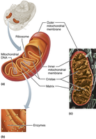
Mitochondria: Double-membraned organelles that produce ATP via aerobic respiration; contain their own DNA and RNA.
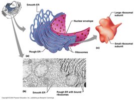
Ribosomes: Sites of protein synthesis; can be free in cytosol or bound to rough ER.
Endoplasmic Reticulum (ER): Network of membranes; rough ER (with ribosomes) synthesizes proteins, smooth ER synthesizes lipids and detoxifies chemicals.
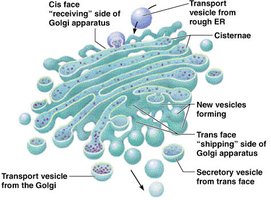
Golgi Apparatus: Modifies, sorts, and packages proteins and lipids for secretion or delivery to other organelles.
Lysosomes: Contain digestive enzymes to break down waste and cellular debris.
Peroxisomes: Contain enzymes for detoxification and neutralization of free radicals.
Cytoskeleton: Network of protein filaments (microfilaments, intermediate filaments, microtubules) that maintain cell shape and facilitate movement.
Centrioles: Organize microtubules during cell division; form bases of cilia and flagella.
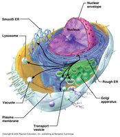
Nucleus and Genetic Material
Nucleus
The nucleus is the control center of the cell, containing the genetic material (DNA) organized as chromatin or chromosomes. It is surrounded by a double membrane (nuclear envelope) with pores for molecular transport.
Nucleoli: Sites of ribosome production within the nucleus.
Chromatin: DNA-protein complex that condenses to form chromosomes during cell division.
Cell Cycle and Division
Phases of the Cell Cycle
The cell cycle consists of interphase (growth and DNA replication) and the mitotic phase (mitosis and cytokinesis). Mitosis ensures equal distribution of genetic material to daughter cells.
Interphase: G1 (growth), S (DNA synthesis), G2 (preparation for division), G0 (non-dividing state).
Mitosis: Prophase, Metaphase, Anaphase, Telophase (PMAT).
Cytokinesis: Division of the cytoplasm, forming two daughter cells.
DNA Replication
DNA replication is semiconservative, producing two identical DNA molecules from one original. Key enzymes include helicase (unwinds DNA), DNA polymerase (synthesizes new strands), and ligase (joins fragments).
Leading Strand: Synthesized continuously.
Lagging Strand: Synthesized in short fragments (Okazaki fragments).
Protein Synthesis
From DNA to Protein
Protein synthesis involves two main processes: transcription (DNA to RNA) and translation (RNA to protein).
Transcription: Synthesis of mRNA from DNA template in the nucleus.
Translation: mRNA is decoded at the ribosome to assemble amino acids into a polypeptide chain.
Types of RNA: mRNA (messenger), tRNA (transfer), rRNA (ribosomal).
Summary Table: Membrane Transport Mechanisms
Process | Energy Source | Description | Example |
|---|---|---|---|
Simple Diffusion | None | Movement of molecules from high to low concentration | Oxygen, CO2 |
Facilitated Diffusion | None | Transport via protein channels or carriers | Glucose, ions |
Osmosis | None | Diffusion of water across membrane | Water movement in/out of cells |
Primary Active Transport | ATP | Pumping of ions against gradient | Na+/K+ pump |
Secondary Active Transport | Ionic gradient | Cotransport of substances | Na+-glucose symport |
Vesicular Transport | ATP | Transport of large particles via vesicles | Endocytosis, exocytosis |
Additional info: This guide covers the essential concepts of cell structure, membrane transport, organelles, and the cell cycle, providing a foundation for further study in anatomy and physiology.
