 Back
Cells: The Living Units – Structure, Function, and Diversity
Study Guide - Smart Notes
 Tailored notes based on your materials, expanded with key definitions, examples, and context.
Cells: The Living Units
Overview of Cells
Cells are the fundamental units of life, forming the basis of all living organisms. The study of cells has been shaped by key scientific discoveries, including those by Robert Hooke, Matthias Schleiden, Theodor Schwann, and Rudolf Virchow. Cells perform essential functions such as obtaining nutrients, synthesizing molecules, disposing of wastes, maintaining shape, and replicating themselves.
Definition: A cell is the smallest structural and functional unit of an organism.
Key Functions: Nutrient uptake, molecule synthesis, waste disposal, shape maintenance, and self-replication.
Components: All cells have a plasma membrane, cytoplasm (with organelles), and a nucleus.
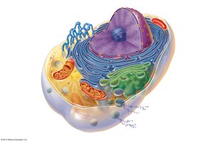
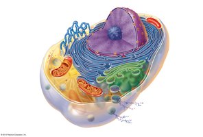
Cellular Organelles and Structures
Major Organelles and Their Functions
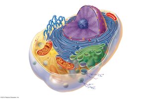
Organelles are specialized subunits within a cell, each with a specific function. Most cells contain the same basic organelles, though their abundance may vary depending on cell type and function.
Nucleus: Control center of the cell; contains DNA and directs protein synthesis.
Plasma Membrane: Defines cell boundaries and regulates the movement of substances in and out.
Cytoplasm: Internal fluid containing organelles and inclusions.
Ribosomes: Sites of protein synthesis; can be free in cytosol or bound to rough ER.
Endoplasmic Reticulum (ER): Rough ER synthesizes proteins; smooth ER synthesizes lipids and detoxifies chemicals.
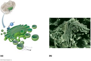
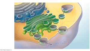
Golgi Apparatus: Modifies, sorts, and packages proteins and lipids for secretion or delivery to other organelles.
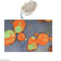
Lysosomes: Contain digestive enzymes to break down waste and cellular debris.
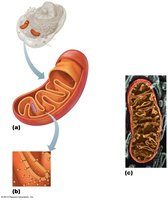
Mitochondria: Generate ATP through cellular respiration; known as the "powerhouse" of the cell.
Peroxisomes: Break down fatty acids and neutralize toxins.
Cytoskeleton: Provides structural support and facilitates cell movement; includes microfilaments, intermediate filaments, and microtubules.
Centrosomes and Centrioles: Organize microtubules and are important in cell division.
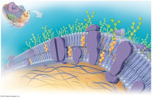
The Plasma Membrane
Structure and Function
The plasma membrane is a selectively permeable barrier that separates the cell's interior from its external environment. It is composed of a phospholipid bilayer with embedded proteins, cholesterol, and carbohydrates.
Fluid Mosaic Model: Describes the dynamic arrangement of lipids and proteins in the membrane.
Integral Proteins: Span the membrane and function as channels, carriers, or receptors.
Peripheral Proteins: Attach to the membrane surface and provide structural support.
Glycocalyx: Carbohydrate-rich area on the cell surface involved in cell recognition and protection.
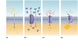
Membrane Transport Mechanisms
Cells regulate the movement of substances across the plasma membrane through various transport mechanisms:
Simple Diffusion: Movement of molecules from high to low concentration without energy input.
Osmosis: Diffusion of water across a selectively permeable membrane.
Facilitated Diffusion: Movement of molecules down their concentration gradient via integral proteins.
Active Transport: Movement of substances against their concentration gradient using ATP and transport proteins.
Endocytosis and Exocytosis
Large particles and macromolecules are transported across the plasma membrane via vesicular transport mechanisms:
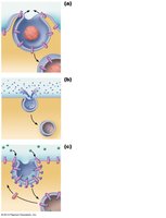
Endocytosis: Uptake of materials into the cell by vesicle formation.
Phagocytosis: "Cell eating" – engulfment of large particles.
Pinocytosis: "Cell drinking" – uptake of extracellular fluid.
Receptor-Mediated Endocytosis: Specific uptake of molecules after binding to receptors.
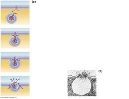
Exocytosis: Release of substances from the cell via vesicle fusion with the plasma membrane.
Cytoplasmic Organelles
Ribosomes
Ribosomes are the sites of protein synthesis, composed of proteins and ribosomal RNA. They may be free in the cytosol or attached to the rough ER. Free ribosomes synthesize proteins for use within the cell, while bound ribosomes produce proteins for secretion or membrane insertion.
Endoplasmic Reticulum (ER)
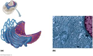
The ER is a network of membranes involved in protein and lipid synthesis. The rough ER is studded with ribosomes and synthesizes proteins, while the smooth ER is involved in lipid metabolism and detoxification.
Golgi Apparatus
The Golgi apparatus modifies, sorts, and packages proteins and lipids received from the rough ER. It is composed of stacked, flattened membranous sacs called cisternae.
Lysosomes
Lysosomes are membrane-bound organelles containing digestive enzymes. They break down unwanted materials and cellular debris, acting as the cell's "demolition crew."
Mitochondria
Mitochondria are double-membraned organelles responsible for producing ATP through cellular respiration. They contain their own DNA and are believed to have originated from ancestral bacteria.
Peroxisomes
Peroxisomes contain enzymes that neutralize free radicals and break down fatty acids and toxins. They are especially abundant in the liver and kidneys.
Cytoskeleton
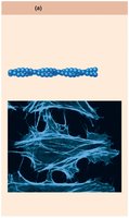
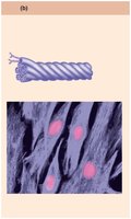
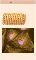
The cytoskeleton is a network of protein filaments that provides structural support, maintains cell shape, and facilitates movement. It consists of:
Microfilaments: Thin filaments of actin involved in cell movement and shape.
Intermediate Filaments: Provide mechanical support and help maintain cell integrity.
Microtubules: Hollow tubes that help with organelle movement and cell division.
Centrosomes and Centrioles
The centrosome is a region near the nucleus that organizes microtubules and contains a pair of centrioles, which are important for cell division and the formation of cilia and flagella.
Cytoplasmic Inclusions
These are temporary structures in the cytoplasm, such as pigment granules, crystals, and food stores (e.g., lipid droplets and glycogen granules).
The Nucleus
Structure and Function
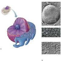
The nucleus is the control center of the cell, containing DNA and directing all cellular activities. It is surrounded by a double membrane called the nuclear envelope, which contains nuclear pores for molecular exchange.
Nucleolus: Site of ribosome subunit assembly.
Chromatin: DNA-protein complex that condenses to form chromosomes during cell division.
Chromatin and Chromosomes
DNA Organization
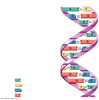
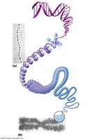
DNA is organized into chromatin, which consists of DNA wrapped around histone proteins to form nucleosomes. Chromatin can be extended (active) or condensed (inactive). During cell division, chromatin condenses further to form chromosomes.
DNA Subunits: Adenine (A), Thymine (T), Cytosine (C), Guanine (G).
Chromosomes: Highest level of DNA organization; humans have 46 chromosomes per cell.
The Cell Life Cycle
Interphase
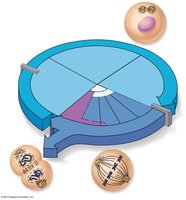
The cell life cycle includes periods of growth, DNA replication, and division. Interphase is divided into three phases:
G1 Phase: Cell growth and normal metabolic activities; centrioles begin to replicate.
S Phase: DNA replication, ensuring genetic continuity.
G2 Phase: Final preparations for division; centrioles finish replicating.
Mitosis and Cytokinesis
Mitosis is the process of nuclear division, followed by cytokinesis, which divides the cytoplasm, resulting in two genetically identical daughter cells. The stages of mitosis are:
Prophase: Chromatin condenses into chromosomes; mitotic spindle forms.
Metaphase: Chromosomes align at the cell's equator.
Anaphase: Sister chromatids separate and move to opposite poles.
Telophase: Chromosomes decondense; nuclear envelopes reform; cytokinesis completes cell division.
Cellular Diversity
Specialized Cell Types
Cells vary in shape, size, and function, reflecting their specialized roles in the body:
Fibroblasts: Synthesize and secrete fibers for connective tissue.
Epithelial Cells: Form linings and barriers; tightly packed for protection.
Erythrocytes (Red Blood Cells): Biconcave shape maximizes surface area for gas exchange.
Muscle Cells: Elongated cells filled with contractile proteins for movement.
Fat Cells: Store nutrients in the form of fat droplets.
Macrophages: Move through tissues to engulf pathogens and debris.
Neurons: Specialized for information transmission and processing.
Sperm Cells: Motile cells specialized for fertilization.
