 Back
BackCellular Anatomy & Physiology: Structure and Function of Cells
Study Guide - Smart Notes
 Tailored notes based on your materials, expanded with key definitions, examples, and context.
Cell Theory and Overview
Introduction to Cell Theory
The cell is the fundamental structural and functional unit of life. All organismal functions depend on the activities of individual and collective cells, and the biochemical activities of cells are dictated by their shapes and specific subcellular structures. Human bodies contain over 200 different types of cells, each specialized for distinct functions.
Cell: The smallest unit of life, capable of performing all vital physiological processes.
Cell Theory: All living things are composed of cells; cells are the basic unit of structure and function; all cells arise from pre-existing cells.
Major Components of the Cell
Three Main Parts of a Cell
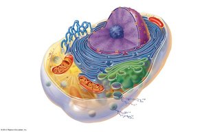
Cells are composed of three primary components: the plasma membrane, cytoplasm, and nucleus. Each part plays a critical role in maintaining cellular integrity and function.
Plasma Membrane: Defines the cell boundary, regulates transport, and facilitates communication.
Cytoplasm: Gel-like substance housing organelles and facilitating metabolic reactions.
Nucleus: Stores genetic material and controls cellular activities.
Plasma Membrane Structure and Function
Fluid Mosaic Model
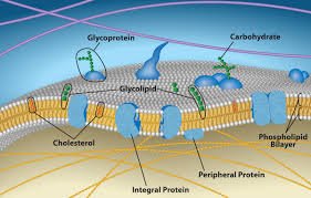
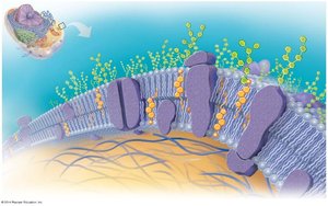
The plasma membrane is described by the fluid mosaic model, consisting of a dynamic lipid bilayer with embedded proteins and cholesterol. This structure allows selective permeability, communication, and structural support.
Lipids: Phospholipids form the bilayer, cholesterol stabilizes fluidity.
Proteins: Integral and peripheral proteins serve as channels, receptors, and anchors.
Carbohydrates: Glycoproteins and glycolipids form the glycocalyx for cell recognition.
Key Functions of the Plasma Membrane
Selective Permeability: Controls entry and exit of substances.
Physical Barrier: Separates intracellular and extracellular environments.
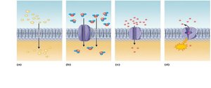
Cell Signaling: Receptors detect and transmit signals.
Cell Recognition: Glycocalyx enables immune recognition.
Structural Support: Anchors cytoskeleton and maintains shape.
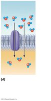
Endocytosis/Exocytosis: Transports large materials via vesicles.
Cytoplasm and Cytosol
Distinction Between Cytoplasm and Cytosol
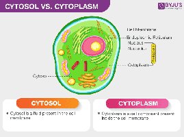
The cytoplasm encompasses all contents within the cell membrane except the nucleus, including cytosol, organelles, and cytoskeleton. Cytosol is the water-based, jelly-like fluid component that surrounds the organelles.
Cytoplasm: Entire content inside the cell membrane (excluding nucleus).
Cytosol: Fluid portion of the cytoplasm.
Functions of Cytoplasm
Structural Support: Maintains cell shape and firmness.
Organelle Suspension: Holds organelles in place.
Metabolic Hub: Site for glycolysis, protein synthesis, and other reactions.
Transport Mechanism: Moves nutrients and proteins.
Waste Management: Contains enzymes for breakdown of waste.
Protection: Buffers genetic material and organelles.
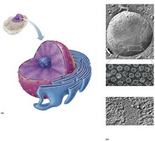
Nucleus
Structure and Function of the Nucleus
The nucleus is the control center of the cell, responsible for storing genetic material, regulating gene expression, and directing cellular activities.
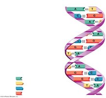
Genetic Storage: Contains DNA, the blueprint for cellular functions.
Control Center: Regulates growth, metabolism, and reproduction.
Gene Expression: Transcribes DNA into mRNA for protein synthesis.
DNA Replication: Ensures genetic continuity during cell division.
Ribosome Synthesis: Nucleolus produces rRNA and assembles ribosomes.
Membrane Transport Mechanisms
Types of Membrane Transport
Substances cross the plasma membrane via passive or active processes, depending on energy requirements and concentration gradients.
Passive Transport: No energy required; includes diffusion, facilitated diffusion, osmosis, and filtration.
Active Transport: Requires ATP; includes primary and secondary active transport, and vesicular transport.
Passive Transport
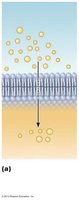
Simple Diffusion: Movement of small, nonpolar molecules down their concentration gradient.
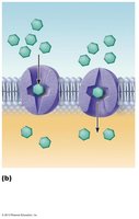
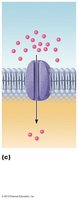
Facilitated Diffusion: Movement of larger or charged molecules via protein carriers or channels.
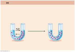
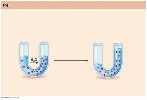
Osmosis: Diffusion of water through aquaporins or the lipid bilayer.
Filtration: Movement across capillary walls due to pressure gradients.
Osmosis and Tonicity
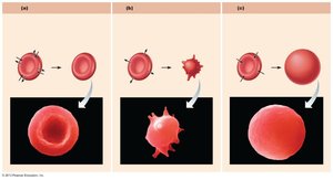
Osmosis affects cell volume and function, with solutions classified as isotonic, hypertonic, or hypotonic based on solute concentration.
Isotonic: Equal solute concentration; no net water movement.
Hypertonic: Higher solute concentration outside; cell shrinks.
Hypotonic: Lower solute concentration outside; cell swells and may lyse.
Active Transport
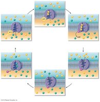
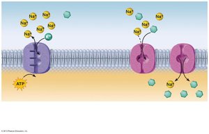
Primary Active Transport: Direct use of ATP to move substances against their gradient (e.g., Na+/K+ pump).
Secondary Active Transport: Indirect use of ATP via ion gradients created by primary transport; includes symport and antiport systems.
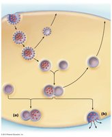
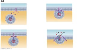
Vesicular Transport: Movement of large particles via vesicles (endocytosis, exocytosis, phagocytosis, pinocytosis).
Cellular Organelles
Major Organelles and Their Functions
Organelles are specialized structures within the cytoplasm that perform distinct functions necessary for cell survival and activity.
Nucleus: Stores genetic material and directs cellular activities.
Ribosomes: Synthesize proteins.
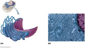
Endoplasmic Reticulum (ER): Smooth ER synthesizes lipids; rough ER synthesizes proteins.
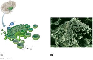
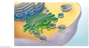
Golgi Apparatus: Modifies, sorts, and packages proteins and lipids.
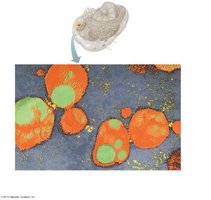
Lysosomes: Digest cellular waste and foreign material.
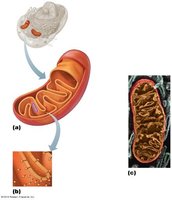
Mitochondria: Produce ATP via cellular respiration.
Peroxisomes: Break down fatty acids and detoxify harmful substances.
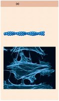
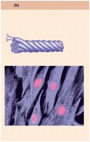
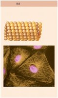
Cytoskeleton: Provides structural support and facilitates movement (microfilaments, intermediate filaments, microtubules).
Nucleus and Genetic Material
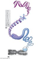
Nuclear Structure and Chromatin Organization
The nucleus contains chromatin (DNA and proteins), the nucleolus, and is surrounded by a nuclear envelope with pores for molecular transport.
Chromatin: DNA wrapped around histone proteins, condenses into chromosomes during cell division.
Nucleolus: Site of ribosome synthesis.
Nuclear Envelope: Double membrane with pores for transport.
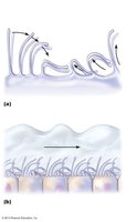
Cellular Extensions
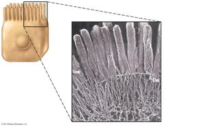
Cilia, Flagella, and Microvilli
Cells may possess extensions that aid in movement or increase surface area for absorption.
Cilia: Motile extensions that move substances across cell surfaces.
Flagella: Longer extensions that propel entire cells (e.g., sperm).
Microvilli: Fingerlike projections that increase surface area for absorption.
Cell Cycle and Division
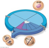
Phases of the Cell Cycle
The cell cycle consists of interphase (G1, S, G2) and the mitotic phase (M), which includes mitosis and cytokinesis. This cycle ensures growth, DNA replication, and division into two daughter cells.
G1 Phase: Cell growth and protein synthesis.
S Phase: DNA replication.
G2 Phase: Preparation for division.
M Phase: Mitosis (nuclear division) and cytokinesis (cytoplasmic division).
Aging and Cellular Lifespan
Theories of Cellular Aging
Aging is a complex process influenced by genetic and environmental factors. Theories include:
Free Radical Theory: Accumulation of metabolic by-products damages cellular molecules.
Mitochondrial Theory: Decreased mitochondrial energy production leads to cellular aging.
Genetic Theory: Aging is programmed by genes; telomeres limit cell division, and telomerase maintains telomere length.
----------------------------------------
