 Back
Cellular Form and Function: Structure, Membrane, and Transport
Study Guide - Smart Notes
 Tailored notes based on your materials, expanded with key definitions, examples, and context.
Cellular Form and Function
Concepts of Cellular Structure
The study of cells, known as cytology, is foundational to understanding anatomy and physiology. The cell theory states that all living organisms are composed of cells, which are the basic structural and functional units of life. Cells arise only from preexisting cells, and an organism’s structure and function are determined by the activities of its cells.
Cell Theory: All organisms are made of cells; cells are the simplest units of life; all cells come from preexisting cells.
Cytology: The scientific study of cells.
Cell Shapes and Sizes
Human cells exhibit a variety of shapes and sizes, each adapted to specific functions. There are about 200 different types of cells in the human body, and their morphology often relates to their role in tissues and organs.
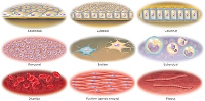
Common Cell Shapes: Squamous (flat), cuboidal (cube-shaped), columnar (tall), polygonal (irregular), stellate (star-shaped), spheroid/ovoid (round/oval), discoidal (disc-shaped), fusiform (spindle-shaped), fibrous (threadlike).
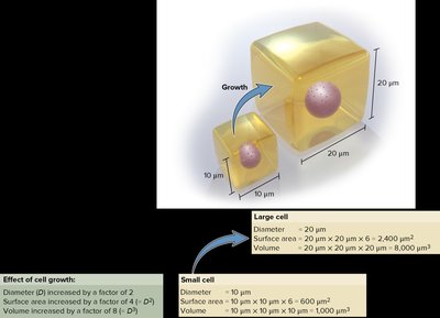
Size Range: Most human cells are 10–15 μm in diameter; egg cells are about 100 μm, and some nerve cells can be over 1 meter long.
Surface Area to Volume Ratio: As a cell grows, its volume increases faster than its surface area, limiting cell size due to the need for adequate exchange with the environment.
Basic Components of a Cell
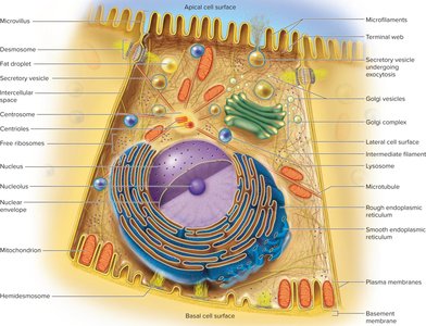
Advances in microscopy have revealed the ultrastructure of cells, including the plasma membrane, cytoplasm, and various organelles. The plasma membrane defines the cell boundary, while the cytoplasm contains organelles, cytoskeleton, inclusions, and cytosol. The extracellular fluid (ECF) surrounds cells and includes interstitial fluid, blood plasma, lymph, and cerebrospinal fluid.
Plasma Membrane: Composed of proteins and lipids; separates the cell from its environment.
Cytoplasm: Contains organelles, cytoskeleton, inclusions, and cytosol (intracellular fluid).
Extracellular Fluid (ECF): Fluid outside the cell, including interstitial fluid and plasma.
The Cell Surface
The Plasma Membrane
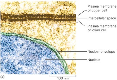
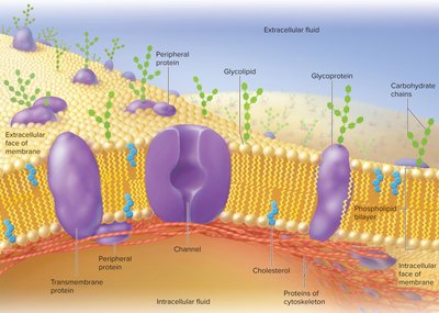
The plasma membrane is a selectively permeable barrier that defines the cell’s boundaries. It consists mainly of a phospholipid bilayer with embedded proteins and carbohydrates.
Phospholipids: Amphipathic molecules forming a bilayer; hydrophilic heads face water, hydrophobic tails face inward.
Cholesterol: Stabilizes membrane fluidity and structure.
Glycolipids: Contribute to the glycocalyx, a carbohydrate coating on the cell surface.
Membrane Proteins
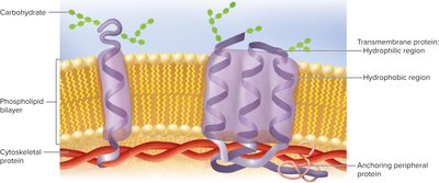
Membrane proteins are crucial for cell function, making up about 2% of the molecules but 50% of the membrane’s weight. They can be transmembrane (spanning the membrane) or peripheral (attached to one side).
Transmembrane Proteins: Span the membrane; involved in transport and signaling.
Peripheral Proteins: Attached to one side; often linked to the cytoskeleton.
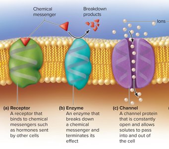
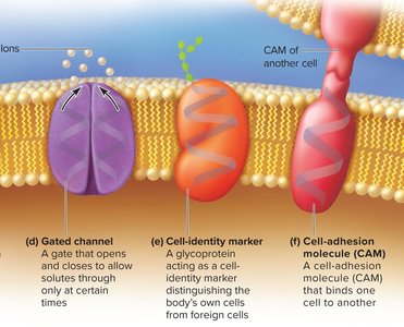
Functions: Receptors, enzymes, channels, carriers, cell-identity markers, and cell-adhesion molecules (CAMs).
Second Messenger Systems
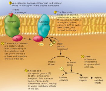
Second messenger systems transmit signals from the cell surface to the interior. For example, when epinephrine binds to a receptor, it activates a G protein, which then triggers the production of cyclic AMP (cAMP) from ATP. cAMP activates kinases, leading to various cellular responses.
First Messenger: The extracellular signal molecule (e.g., hormone).
Second Messenger: Intracellular signaling molecule (e.g., cAMP).
G Proteins: Relay signals from receptors to enzymes.
The Glycocalyx
The glycocalyx is a carbohydrate-rich layer on the cell surface, unique to each individual (except identical twins). It plays roles in protection, immunity, cell adhesion, fertilization, and embryonic development.
Functions: Protection, immunity, defense against cancer, transplant compatibility, cell adhesion, fertilization, embryonic development.
Extensions of the Cell Surface
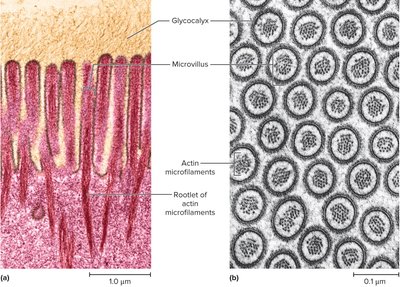
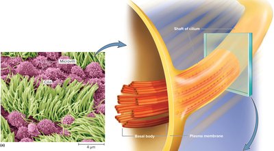
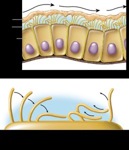
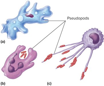
Cells may have surface extensions such as microvilli, cilia, flagella, and pseudopods, each serving specialized functions.
Microvilli: Increase surface area for absorption; contain actin filaments.
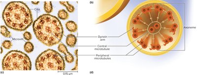
Cilia: Hair-like structures for movement of substances; motile cilia have a 9+2 microtubule arrangement.
Flagellum: Whip-like structure for cell movement (e.g., sperm tail).
Pseudopods: Temporary extensions for movement and phagocytosis.
Membrane Transport
Selective Permeability
The plasma membrane is selectively permeable, allowing some substances to pass while restricting others. Transport mechanisms are classified as passive (no ATP required) or active (require ATP).
Passive Mechanisms: Filtration, diffusion, osmosis.
Active Mechanisms: Active transport, vesicular transport.
Carrier-Mediated Mechanisms: Use membrane proteins to transport substances.
Filtration
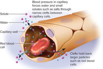
Filtration is the movement of particles through a membrane by physical pressure, such as the filtration of water and solutes through capillary walls in tissues and kidneys.
Simple Diffusion
Simple diffusion is the net movement of particles from high to low concentration due to random molecular motion. It does not require energy and occurs down a concentration gradient.
Factors Affecting Diffusion Rate: Temperature, molecular weight, concentration gradient steepness, membrane surface area, membrane permeability.
Osmosis
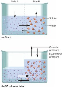
Osmosis is the net flow of water across a selectively permeable membrane from an area of higher water concentration to lower water concentration. Aquaporins facilitate water movement. Osmotic imbalances can cause clinical issues such as edema or dehydration.
Osmolarity and Tonicity
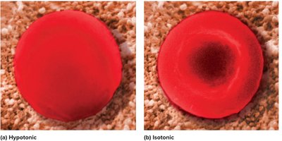
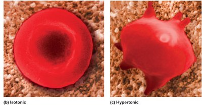
Osmolarity is the total concentration of nonpermeating solutes in a solution. Tonicity describes how a solution affects cell volume:
Hypotonic: Lower solute concentration outside; cell swells and may burst.
Hypertonic: Higher solute concentration outside; cell shrivels.
Isotonic: Equal solute concentration; no net water movement.
Carrier-Mediated Transport
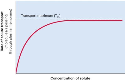
Carrier proteins in the membrane transport solutes by binding and releasing them on opposite sides. They exhibit specificity and saturation (transport maximum, Tm).
Types of Carriers: Uniport (one solute), symport (cotransport, same direction), antiport (countertransport, opposite directions).
Mechanisms: Facilitated diffusion (down gradient, no ATP), primary active transport (up gradient, uses ATP), secondary active transport (uses ATP indirectly).
Example: The sodium–potassium pump (Na+–K+ ATPase) is a primary active transport mechanism that maintains cellular ion gradients, regulates cell volume, and contributes to membrane potential.
*Additional info: The notes above expand on the original content by providing definitions, examples, and clinical relevance where appropriate, ensuring a comprehensive and self-contained study guide for college-level anatomy and physiology students.*
