 Back
BackCellular Level of Organization: Structure and Function of Cells and Their Organelles
Study Guide - Smart Notes
 Tailored notes based on your materials, expanded with key definitions, examples, and context.
Introduction to Cells
Cell Theory and Cellular Differentiation
The cell theory is a fundamental concept in biology, stating that all living organisms are composed of cells, cells are the basic units of structure and function in living things, and all cells arise from pre-existing cells. Cellular differentiation is the process by which a less specialized cell becomes a more specialized cell type, allowing for the diversity of cell functions in multicellular organisms.
Body Fluid Distribution and Cell Structure
Body Fluid Compartments
Extracellular fluid (ECF): Fluid outside cells, including plasma and interstitial fluid.
Interstitial fluid: Fluid between cells within tissues.
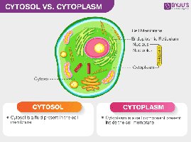
Intracellular fluid (cytosol): Fluid within cells, containing dissolved ions, nutrients, and proteins.
Basic Cell Structure
Plasma membrane: Outer boundary of the cell, separating the internal environment from the external environment.
Cytoplasm: Material within the cell membrane, excluding the nucleus; consists of cytosol and organelles.
Cell Organelles and Their Functions
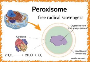
Peroxisome
Structure: Small, membrane-bound vesicles containing enzymes.
Function: Break down hydrogen peroxide (H2O2) into water and oxygen, detoxifying harmful substances. They contain catalase and other oxidative enzymes.
Equation:
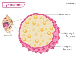
Lysosome
Structure: Membrane-bound vesicles containing hydrolytic enzymes.
Function: Digest and recycle cellular waste, damaged organelles, and foreign materials through enzymatic breakdown.
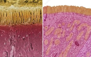
Microvilli
Structure: Membrane extensions containing microfilaments, increasing surface area.
Function: Enhance absorption and secretion by increasing the cell's surface area.
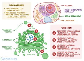
Golgi Apparatus
Structure: Stacks of flattened membranous sacs (cisternae).
Function: Modifies, sorts, and packages proteins and lipids for secretion or delivery to other organelles.
Nucleus
Structure: Surrounded by a double membrane (nuclear envelope), contains nucleoplasm, DNA, nucleotides, and enzymes.
Function: Stores genetic information, controls gene expression, and regulates cell activities.
Nucleolus
Structure: Dense region within the nucleus.
Function: Site of ribosomal RNA (rRNA) synthesis and ribosome assembly.
Endoplasmic Reticulum (ER)
Structure: Network of membranous sheets and channels.
Smooth ER: Lacks ribosomes; synthesizes lipids, detoxifies chemicals, and stores calcium ions.
Rough ER: Studded with ribosomes; synthesizes proteins for secretion or membrane insertion.

Ribosomes
Structure: Composed of RNA and proteins; can be free in cytosol or bound to rough ER.
Function: Synthesize proteins by translating messenger RNA (mRNA).
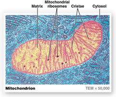
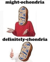
Mitochondrion
Structure: Double-membraned organelle; inner membrane forms cristae and contains metabolic enzymes.
Function: Site of aerobic respiration and ATP production, often called the "powerhouse of the cell." Mitochondria require oxygen and produce carbon dioxide as a byproduct.
Cytoskeleton
Structure: Network of protein filaments and tubules (microfilaments, intermediate filaments, microtubules).
Function: Provides structural support, maintains cell shape, and facilitates intracellular transport and cell division.
The Plasma Membrane
Structure and Function
The plasma membrane is a selectively permeable barrier that separates the cell's interior from the extracellular environment. It regulates the entry and exit of ions, nutrients, and waste products, and is involved in cell signaling and adhesion.
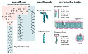
Phospholipid bilayer: Two layers of amphipathic phospholipids with hydrophilic heads facing outward and hydrophobic tails inward.
Cholesterol: Interspersed within the bilayer, stabilizing membrane fluidity.
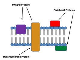
Proteins: Integral (span the membrane), transmembrane, and peripheral (attached to one side).
Glycocalyx: Carbohydrate-rich coating composed of glycoproteins, glycolipids, and proteoglycans, important for cell recognition and protection.
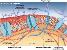
Membrane Proteins and Their Functions
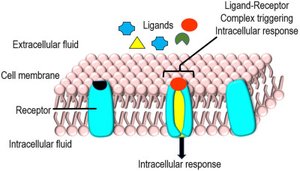
Receptor proteins: Bind specific molecules (ligands) and trigger cellular responses.
Carrier proteins: Transport specific substances across the membrane.
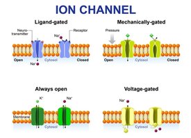
Channels: Allow passage of water and small ions; can be gated or always open.
Review Questions and Key Concepts
Which structural component of the plasma membrane is mostly responsible for isolating a cell from its external environment? The phospholipid bilayer.
General functions of the plasma membrane: Physical isolation, regulation of exchange, sensitivity to the environment, structural support.
Which type of integral protein allows water and small ions to pass through the plasma membrane? Channel proteins.
What characteristic of phospholipids accounts for their packing into a double layer? Their amphipathic nature (hydrophilic heads and hydrophobic tails).
Mitochondria: The Powerhouse of the Cell
Structure and Significance
Mitochondria are double-membraned organelles responsible for producing most of the cell's ATP through aerobic respiration. The presence of many mitochondria in a cell indicates high energy requirements. Oxygen is required for ATP production, and carbon dioxide is produced as a waste product.

Equation for aerobic respiration:
Additional info: The mitochondria contain their own DNA and ribosomes, supporting the endosymbiotic theory of their origin.
