 Back
BackCellular Level of Organization: Structure, Function, and Processes
Study Guide - Smart Notes
 Tailored notes based on your materials, expanded with key definitions, examples, and context.
The Cellular Level of Organization
Protein Synthesis
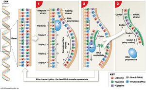
Protein synthesis is a fundamental cellular process by which cells assemble functional polypeptides, determining cell structure and function. This process involves gene activation, transcription, and translation.
Gene Activation: DNA is uncoiled and histones are temporarily removed to expose the gene for transcription.
Transcription: The synthesis of RNA from a DNA template. RNA polymerase binds to the promoter region, reads the DNA code, and links nucleotides to form messenger RNA (mRNA) in codons. The coding strand of DNA specifies the amino acid sequence, while the template strand is used for mRNA production.
RNA Processing: Before mRNA leaves the nucleus, noncoding sequences (introns) are removed and coding segments (exons) are spliced together.
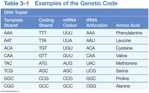
Genetic Code
The genetic code is the set of rules by which information encoded in mRNA is translated into proteins. Each mRNA codon corresponds to a specific amino acid, delivered by transfer RNA (tRNA).
DNA Triplet | Coding Strand | mRNA Codon | tRNA Anticodon | Amino Acid |
|---|---|---|---|---|
AAA | TTT | UUU | AAA | Phenylalanine |
AAT | TTA | UUA | AAU | Leucine |
ACA | TGT | UGU | ACA | Cysteine |
CAA | GTT | GUU | CAA | Valine |
TAC | ATG | AUG | UAC | Methionine |
TCG | AGC | AGC | UCG | Serine |
GCG | CGC | CGC | GCG | Proline |
CGG | GCC | GCC | CGG | Alanine |
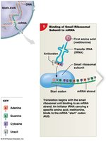
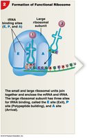
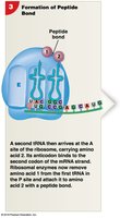
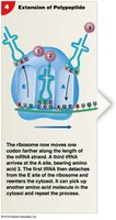
Translation
Translation is the process by which mRNA is decoded to build a polypeptide chain. Ribosomes read mRNA codons, and tRNA delivers the corresponding amino acids, which are joined by peptide bonds. The process ends at a stop codon.
Initiation: mRNA binds to ribosomal subunits in the cytoplasm.
Elongation: tRNA anticodons pair with mRNA codons, and amino acids are linked.
Termination: At the stop codon, the components separate, releasing the completed polypeptide.

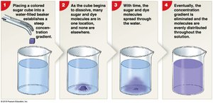
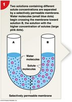
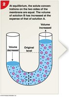
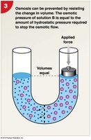
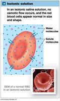
Diffusion and Osmosis
Diffusion and osmosis are passive transport processes essential for cellular homeostasis. The plasma membrane's selective permeability allows cells to regulate the movement of substances.
Diffusion: Net movement of molecules from high to low concentration, driven by random motion and concentration gradients.
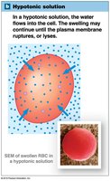
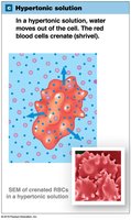
Osmosis: Diffusion of water across a selectively permeable membrane toward higher solute concentration.
Osmotic Pressure: The force required to prevent water movement due to solute concentration differences.
Tonicity: Describes how a solution affects cell volume (isotonic, hypotonic, hypertonic).
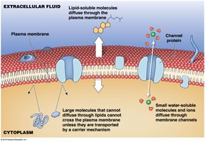
Carrier-Mediated and Vesicular Transport
Cells use carrier proteins and vesicles to transport substances across the plasma membrane, either passively or actively.
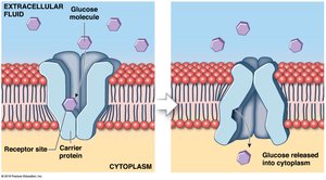
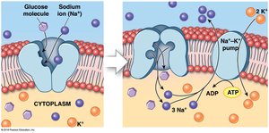
Carrier-Mediated Transport: Proteins transport ions or organic substrates with specificity and saturation limits. Includes facilitated diffusion (passive) and active transport (requires ATP).
Facilitated Diffusion: Carrier proteins help large molecules (e.g., glucose) cross the membrane.
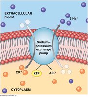
Active Transport: Proteins move substrates against concentration gradients, such as the sodium-potassium exchange pump.
Secondary Active Transport: Uses ATP to establish a gradient for one substance, which then drives the transport of another.
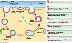
Vesicular Transport: Bulk movement of materials via vesicles, including endocytosis (receptor-mediated, pinocytosis, phagocytosis) and exocytosis.
Membrane Potential
Membrane potential is the electrical potential difference across the plasma membrane, resulting from the separation of positive and negative charges. It is crucial for nerve impulse transmission and muscle contraction.
Resting Membrane Potential: Typically ranges from −10 mV to −100 mV, depending on cell type.
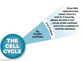
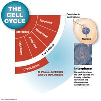
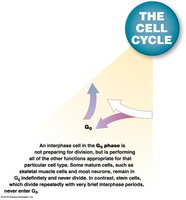
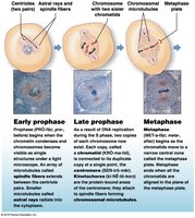
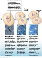
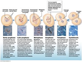
Cell Life Cycle
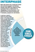
The cell life cycle includes interphase, mitosis, and cytokinesis, which are essential for growth, repair, and maintenance of tissues.
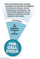
Interphase: Nondividing period with G0, G1, S, and G2 phases. Most somatic cells spend the majority of their lives in interphase.
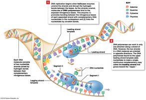
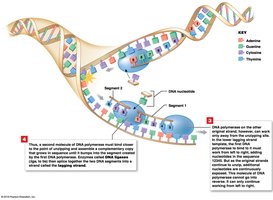
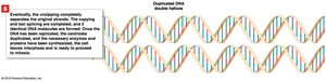
DNA Replication: Helicases unwind DNA, and DNA polymerase synthesizes new strands by bonding complementary nucleotides.
Mitosis: Division of the nucleus into two identical sets of chromosomes, followed by cytokinesis (division of cytoplasm).
Mitotic Rate: The rate of cell division; slower rates mean longer cell life. Muscle cells and neurons rarely divide, while exposed cells are frequently replaced.
Regulation of the Cell Life Cycle
Cell division is tightly regulated to balance cell loss and growth. Internal and external factors can stimulate or inhibit division.
Stimulatory Factors: M-phase promoting factor (MPF), growth factors.
Inhibitory Factors: Repressor genes, worn out telomeres.
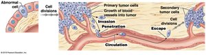
Cell Division and Cancer
Cancer arises from abnormal cell proliferation due to mutations in genes controlling cell growth. Tumors may be benign (contained) or malignant (invasive and metastatic).
Oncogenes: Modified genes that promote cancer.
Mutagens: Agents causing mutations; carcinogens are mutagens that cause cancer.
Metastasis: Spread of cancer to other tissues, beginning with invasion.
Cellular Differentiation
Cellular differentiation is the process by which cells become specialized by turning off genes not needed for their specific function. This allows for the formation of diverse cell types such as liver cells, fat cells, and neurons.
Importance: Enables multicellular organisms to develop tissues and organs with specialized functions.
Key Equations
Concentration Gradient (Diffusion): Where J is the flux, D is the diffusion coefficient, and \frac{dC}{dx} is the concentration gradient.
Osmotic Pressure: Where \Pi is osmotic pressure, i is the van 't Hoff factor, M is molarity, R is the gas constant, and T is temperature in Kelvin.
Summary Table: Types of Membrane Transport
Type | Energy Required | Direction | Example |
|---|---|---|---|
Simple Diffusion | No | High to Low | O2, CO2 |
Facilitated Diffusion | No | High to Low | Glucose |
Active Transport | Yes (ATP) | Low to High | Na+/K+ pump |
Osmosis | No | Water: High to Low | Water movement |
Endocytosis | Yes (ATP) | Into cell | Phagocytosis |
Exocytosis | Yes (ATP) | Out of cell | Secretion |
Additional info: These notes expand on the original content with definitions, examples, and equations to provide a comprehensive, self-contained study guide for college-level anatomy and physiology students.
