 Back
BackCentral and Peripheral Nervous System: Structure and Function
Study Guide - Smart Notes
 Tailored notes based on your materials, expanded with key definitions, examples, and context.
The Central Nervous System (CNS)
Overview of the CNS
The central nervous system consists of the brain and spinal cord, serving as the main control center for the body. It processes sensory information, coordinates voluntary and involuntary responses, and integrates higher cognitive functions.
Brain: Divided into four major regions: cerebrum, diencephalon, brain stem, and cerebellum.
Spinal Cord: Connects the brain to the peripheral nervous system and mediates reflexes.
Major Regions of the Brain
Cerebrum: Largest part, responsible for higher brain functions such as thought, action, and sensory processing.
Diencephalon: Contains the thalamus and hypothalamus, relaying sensory information and regulating homeostasis.
Brain Stem: Includes midbrain, pons, and medulla oblongata; controls basic life functions.
Cerebellum: Coordinates movement and balance.
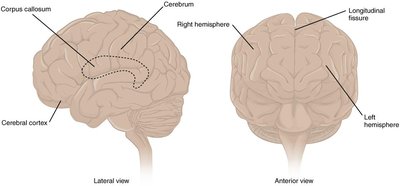
Cerebrum Structure
The cerebrum is characterized by its wrinkled outer layer, the cerebral cortex, composed of grey matter (neuron cell bodies and dendrites). The surface features gyri (ridges) and sulci (depressions), which increase surface area for higher cognitive functions.
Longitudinal fissure: Deep groove dividing the left and right cerebral hemispheres.
Corpus callosum: White matter tract connecting the hemispheres, allowing communication between them.
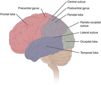
Lobes of the Cerebral Cortex
The cerebral cortex is divided into four lobes, each associated with specific functions:
Frontal lobe: Higher cognitive functions (decision making, problem-solving), motor cortex (voluntary movement), prefrontal cortex (personality, intelligence).
Parietal lobe: Processes tactile senses (touch, pressure, pain), proprioception, and visual perception.
Occipital lobe: Visual processing.
Temporal lobe: Auditory processing, language comprehension.
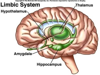
Subcortical Structures: The Limbic System
Located deep to the cerebral cortex, the limbic system is the center of emotional and behavioral expression. It includes the amygdala, hippocampus, and hypothalamus.
Amygdala: Involved in fear, anxiety, and long-term memory formation.
Hippocampus: Essential for long-term memory formation.
Hypothalamus: Regulates memory, emotion, and homeostasis (body temperature, circadian rhythm, food/fluid intake, autonomic nervous system).
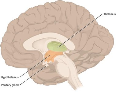
Diencephalon
The diencephalon, located beneath the cerebrum, is primarily composed of the thalamus and hypothalamus. It acts as a relay center for sensory and motor signals between the cerebrum and the rest of the nervous system.
Thalamus: Principal relay for all sensory information (except olfaction), processes and propagates sensory input to the cerebrum.
Hypothalamus: Major control center for homeostasis and endocrine regulation via the pituitary gland.
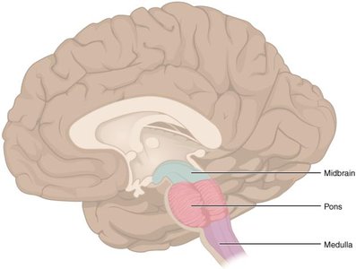
Brain Stem
The brain stem consists of the midbrain, pons, and medulla oblongata. It contains major ascending (sensory) and descending (motor) tracts connecting the cerebrum and spinal cord.
Midbrain: Processes auditory and visual information; contains nuclei for cranial nerves III and IV.
Pons: Bridge between cerebellum and brain stem; contains nuclei for cranial nerves V–VIII.
Medulla oblongata: Regulates heart rate, blood pressure, and breathing; contains nuclei for cranial nerves IX–XII.
Cerebellum
The cerebellum, or "little brain," has gyri and sulci similar to the cerebrum. It is crucial for maintaining balance, posture, and coordinating voluntary movements by comparing motor commands with sensory feedback.
Spinal Cord Structure
The spinal cord is composed of grey and white matter. Grey matter is organized into horns:
Anterior horns: Motor neuron cell bodies; send signals to skeletal muscles.
Posterior horns: Receive sensory information from the body.
Lateral horns: (Thoracic/upper lumbar only) Visceral motor neuron cell bodies; part of the autonomic nervous system.
White matter is organized into columns containing ascending (sensory) and descending (motor) tracts.
Meninges and Cerebrospinal Fluid (CSF)
The CNS is protected by three connective tissue membranes called meninges:
Dura mater: Tough, outermost layer attached to the skull.
Arachnoid mater: Middle layer forming a sac around the CNS; subarachnoid space contains CSF.
Pia mater: Thin, delicate layer lining the brain's surface and sulci.
CSF circulates through the ventricular system (lateral, third, and fourth ventricles) and subarachnoid space, providing cushioning and nutrient transport. CSF is produced by ependymal cells and reabsorbed via arachnoid granulations.
The Peripheral Nervous System (PNS)
Ganglia and Nerves
A ganglion is a cluster of neuron cell bodies in the PNS, classified as sensory or autonomic. The most common sensory ganglia are dorsal root ganglia, containing pseudo-unipolar neurons that transmit sensory information to the CNS.
Nerves are bundles of axons in the PNS, surrounded by connective tissue layers:
Epineurium: Surrounds the entire nerve.
Perineurium: Surrounds bundles (fascicles) of axons.
Endoneurium: Surrounds individual axons.
Spinal Nerves
There are 31 pairs of spinal nerves, named for the spinal cord region from which they emerge. Each spinal nerve splits into a dorsal (sensory) and ventral (motor) root near the spinal cord. Many pathways are contralateral, meaning sensory/motor information crosses to the opposite side of the brain.
Cranial Nerves
There are 12 pairs of cranial nerves (CN I–XII), primarily responsible for sensory and motor functions of the head and neck. Each nerve has specific functions:
CN I (Olfactory): Sensory, smell
CN II (Optic): Sensory, vision
CN III (Oculomotor): Motor, eye movement, eyelid, pupil constriction
CN IV (Trochlear): Motor, eye movement
CN V (Trigeminal): Sensory and motor, facial sensation, mastication
CN VI (Abducens): Motor, eye movement
CN VII (Facial): Sensory and motor, facial expression, taste, saliva production
CN VIII (Vestibulocochlear): Sensory, hearing, balance
CN IX (Glossopharyngeal): Sensory and motor, taste, swallowing, saliva production
CN X (Vagus): Sensory and motor, swallowing, voice, autonomic control of thoracic/abdominal organs
CN XI (Accessory): Motor, swallowing, head/neck/shoulder movement
CN XII (Hypoglossal): Motor, tongue movement
Summary Table: Major Brain Regions and Functions
Region | Main Structures | Primary Functions |
|---|---|---|
Cerebrum | Cerebral cortex, corpus callosum | Higher cognitive functions, sensory/motor processing |
Diencephalon | Thalamus, hypothalamus | Sensory relay, homeostasis, endocrine control |
Brain Stem | Midbrain, pons, medulla | Basic life functions, cranial nerve nuclei |
Cerebellum | Gyri, sulci | Balance, posture, movement coordination |
