 Back
BackChapter 12 Guided Notes
Study Guide - Smart Notes
 Tailored notes based on your materials, expanded with key definitions, examples, and context.
Q1. What is the difference between gray matter and white matter?
Background
Topic: Brain Anatomy – Tissue Organization
This question tests your understanding of the structural and functional differences between gray matter and white matter in the central nervous system.
Key Terms:
Gray Matter: Contains neuron cell bodies, dendrites, and unmyelinated axons.
White Matter: Composed mainly of myelinated axons, which give it a lighter appearance.
Step-by-Step Guidance
Consider what structures are found in gray matter versus white matter (think about cell bodies, axons, and myelin).
Think about the color difference and what causes it (hint: myelin is a fatty substance).
Reflect on where each type is located in the brain and spinal cord, and their primary functions (processing vs. transmission).
Try solving on your own before revealing the answer!
Final Answer:
Gray matter consists mainly of neuron cell bodies, dendrites, and unmyelinated axons, and is involved in processing and integration of information. White matter is made up of myelinated axons, which are responsible for transmitting signals between different brain regions and between the brain and spinal cord. The myelin sheath gives white matter its lighter color.
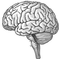
Q2. Identify this view of the brain and label with corresponding list terms. Be sure to label the major sulci and gyri.
Background
Topic: Brain Anatomy – External Landmarks
This question asks you to recognize the orientation of a brain image and identify key external features, including sulci (grooves) and gyri (ridges).
Key Terms:
Gyri: Elevated ridges on the cerebral cortex.
Sulci: Shallow grooves separating gyri.
Lobes: Major regions of the brain (frontal, parietal, temporal, occipital).
Step-by-Step Guidance
Observe the orientation of the brain (is it a lateral, medial, or superior view?).
Identify the major lobes visible in this view (frontal, parietal, occipital, temporal).
Locate and label the central sulcus, precentral gyrus, postcentral gyrus, and lateral sulcus.
Use anatomical landmarks to help you place each label accurately.
Try solving on your own before revealing the answer!
Final Answer:
This is a lateral (external) view of the brain. Major features to label include:
Central sulcus
Precentral gyrus (primary motor cortex)
Postcentral gyrus (primary somatosensory cortex)
Lateral sulcus (Sylvian fissure)
Frontal, parietal, temporal, and occipital lobes
Q3. Elevated ridges on the cerebral surface are called __________, while shallow grooves are __________.
Background
Topic: Brain Surface Anatomy
This question tests your knowledge of the terminology for the surface features of the cerebral cortex.
Key Terms:
Gyri: Elevated ridges
Sulci: Shallow grooves
Step-by-Step Guidance
Recall the terms for the raised and indented features on the brain's surface.
Match the correct term to each description: elevated ridge and shallow groove.
Try solving on your own before revealing the answer!
Final Answer:
Elevated ridges are called gyri, while shallow grooves are sulci.
Q4. Deep grooves separating brain regions are called __________.
Background
Topic: Brain Surface Anatomy
This question focuses on the terminology for deeper grooves in the brain that separate major regions or lobes.
Key Terms:
Fissures: Deep grooves in the brain, such as the longitudinal fissure and transverse fissure.
Step-by-Step Guidance
Recall the term used for grooves deeper than sulci.
Think of examples, such as the longitudinal fissure separating the cerebral hemispheres.
Try solving on your own before revealing the answer!
Final Answer:
Deep grooves separating brain regions are called fissures.
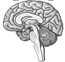
Q5. Identify this view of the brain and label it with corresponding list terms.
Background
Topic: Brain Anatomy – Internal Structures
This question asks you to recognize a mid-sagittal (internal) view of the brain and identify key internal structures.
Key Terms:
Corpus callosum
Thalamus
Hypothalamus
Cerebellum
Brainstem (midbrain, pons, medulla oblongata)
Step-by-Step Guidance
Identify the orientation of the brain (mid-sagittal section).
Locate the corpus callosum, thalamus, hypothalamus, cerebellum, and brainstem.
Label each structure based on its position and shape.
Try solving on your own before revealing the answer!
Final Answer:
This is a mid-sagittal (internal) view of the brain. Key structures to label include:
Corpus callosum
Thalamus
Hypothalamus
Cerebellum
Midbrain, pons, medulla oblongata
Q6. Match the Brain structure to its Function
Background
Topic: Brain Regions and Functions
This question tests your ability to associate specific brain structures with their primary functions.
Key Terms:
Cerebellum: Balance and coordination
Thalamus: Sensory relay station
Hypothalamus: Homeostasis (temperature, hunger, etc.)
Medulla oblongata: Autonomic control (heart rate, respiration)
Frontal lobe: Voluntary movement, decision making
Occipital lobe: Visual processing
Pons: Bridge for signals, breathing regulation
Step-by-Step Guidance
Read each function and recall which brain structure is responsible for it.
Match each function to the correct lettered structure from the list.
Double-check your matches by reviewing the main roles of each brain region.
Try solving on your own before revealing the answer!
Final Answer:
1. G (Occipital lobe – Visual processing center)
2. C (Hypothalamus – Regulation of body temperature and hunger)
3. A (Cerebellum – Coordination of balance and motor control)
4. B (Thalamus – Relay station for sensory information)
5. D (Medulla oblongata – Control of heart rate and respiration)
7. G (Pons – Bridge between brain regions and breathing regulation)
8. E (Frontal lobe – Voluntary motor planning and decision making)
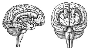
Q7. The images below depict the ventricular system within the brain. Label the four ventricles within each view.
Background
Topic: Brain Ventricular System
This question tests your ability to identify the four ventricles of the brain in different anatomical views.
Key Terms:
Lateral ventricles
Third ventricle
Fourth ventricle
Step-by-Step Guidance
Identify the lateral, third, and fourth ventricles in both the lateral and anterior views.
Label the paired lateral ventricles, the single third ventricle, and the fourth ventricle.
Note the connections between the ventricles (interventricular foramen, cerebral aqueduct).
Try solving on your own before revealing the answer!
Final Answer:
Lateral ventricles (right and left)
Third ventricle
Fourth ventricle
These are the four main ventricles of the brain, visible in both lateral and anterior views.
Q8. List two functions of cerebrospinal fluid.
Background
Topic: CNS Protection and Homeostasis
This question tests your understanding of the roles of cerebrospinal fluid (CSF) in the central nervous system.
Key Terms:
Cerebrospinal fluid (CSF): Clear fluid in the brain and spinal cord
Step-by-Step Guidance
Think about how CSF protects the brain physically (e.g., cushioning, buoyancy).
Consider how CSF helps maintain a stable environment for neural tissue (e.g., nutrient delivery, waste removal).
Try solving on your own before revealing the answer!
Final Answer:
CSF provides buoyancy and shock absorption for the brain and spinal cord.
CSF helps deliver nutrients and remove waste products from the CNS.
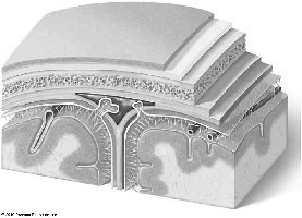
Q9. Meninges: Identify the brain's meningeal layers and associated structures.
Background
Topic: CNS Protection – Meninges
This question asks you to identify the three meningeal layers and related structures that protect the brain.
Key Terms:
Dura mater: Tough outer layer
Arachnoid mater: Web-like middle layer
Pia mater: Thin inner layer, adheres to brain surface
Subarachnoid space: Contains CSF
Step-by-Step Guidance
Identify the three meningeal layers from outermost to innermost.
Label the dura mater, arachnoid mater, and pia mater.
Locate the subarachnoid space and note its contents.
Try solving on your own before revealing the answer!
Final Answer:
Dura mater (outermost)
Arachnoid mater (middle, web-like)
Pia mater (innermost, adheres to brain)
Subarachnoid space (contains CSF)
Q10. Matching: CNS Protection & Support
Background
Topic: CNS Protective Structures
This question tests your ability to match CNS protective/support structures to their functions.
Key Terms:
Dura mater: Tough outer meningeal layer
Arachnoid mater: Web-like middle meningeal layer
Pia mater: Thin, delicate inner meningeal layer
Choroid plexus: Produces CSF
Blood-brain barrier: Limits passage of substances into CNS
Cerebrospinal fluid: Provides buoyancy and shock absorption
Step-by-Step Guidance
Read each function and recall which structure is responsible for it.
Match each function to the correct lettered structure from the list.
Double-check your matches by reviewing the main roles of each structure.
Try solving on your own before revealing the answer!
Final Answer:
1. D (Choroid plexus – Produces cerebrospinal fluid)
2. A (Dura mater – Tough outer meningeal layer)
3. C (Pia mater – Closely adheres to brain surface)
4. B (Arachnoid mater – Web-like middle meningeal layer)
5. E (Blood-brain barrier – Limits passage of harmful substances into CNS)
6. F (Cerebrospinal fluid – Provides buoyancy and shock absorption)
Q11. Cranial Nerves: Identify The 12 Pairs of Cranial Nerves. List each cranial nerve by name, number and determine if each is sensory, motor, or mixed (both).
Background
Topic: Cranial Nerves
This question tests your ability to recall the names, numbers, and functions (sensory, motor, or both) of the 12 cranial nerves.
Key Terms:
Cranial nerves: 12 pairs, each with a specific name, number, and function
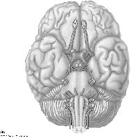
Step-by-Step Guidance
List the cranial nerves in order (I–XII) by their Roman numerals.
Write the name of each nerve next to its number.
Determine if each nerve is primarily sensory, motor, or mixed (both).
Use a mnemonic to help remember the order and function if needed.
Olfactory
Optic
Oculomotor
Trochelar
Trigeminal
Abducens
Facial
Vestibulocochlear
Glossopharyngeal
Vagus
Accessory
Hypoglossal
Try solving on your own before revealing the answer!
Final Answer:
I. Olfactory – Sensory
II. Optic – Sensory
III. Oculomotor – Motor
IV. Trochlear – Motor
V. Trigeminal – Both
VI. Abducens – Motor
VII. Facial – Both
VIII. Vestibulocochlear – Sensory
IX. Glossopharyngeal – Both
X. Vagus – Both
XI. Accessory – Motor
XII. Hypoglossal – Motor
