 Back
BackCentral Nervous System: Brain Structure, Function, and Protection
Study Guide - Smart Notes
 Tailored notes based on your materials, expanded with key definitions, examples, and context.
CNS – The Brain, Part 2
Diencephalon
The diencephalon is a central brain region that acts as a relay and control hub, integrating sensory, motor, and autonomic functions. It includes key structures like the thalamus, hypothalamus, epithalamus, and subthalamus, each with distinct roles in maintaining homeostasis and processing information.
Brainstem
The brainstem is the lower part of the brain that connects to the spinal cord. It consists of three main regions:
Midbrain: Located just below the thalamus. Controls visual and auditory reflexes. Important for motor movement and coordination.
Pons: Middle section of the brainstem. Acts as a bridge between the cerebrum and cerebellum. Regulates breathing and communication between brain regions.
Medulla Oblongata: Lowest part, continuous with the spinal cord. Controls vital autonomic functions like heart rate, blood pressure, and respiration.
Functions of the Brainstem:
Basic life support (breathing, heartbeat)
Reflexes (swallowing, coughing)
Pathway for sensory and motor signals between brain and body
Cerebellum
The cerebellum comprises about 2.5% of total brain mass and is located posteriorly beneath the occipital lobes and behind the brainstem. It has two hemispheres connected by the vermis, with a highly folded surface (folia).
Grey matter: Cerebellar cortex & deep nuclei
White matter: Arbor vitae and cerebellar peduncles
Functions:
Motor Coordination: Fine-tunes voluntary movements
Balance & Posture: Maintains equilibrium
Motor Learning: Important for learning new motor skills
Cognitive Roles: Involved in attention and language processing
Lobes and Structures:
Anterior Lobe: Coordinates posture and limb movements
Posterior Lobe: Fine motor control and voluntary movement
Flocculonodular Lobe: Maintains balance and eye movements
Vermis: Connects the two hemispheres and helps with posture control
Functional Brain Systems
Limbic System
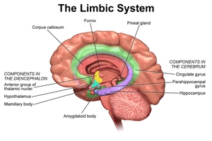
The limbic system is a network of structures involved in emotion, memory, and learning. It includes:
Amygdala: Expression of emotion, arousal, fear; association between a stimulus and its emotional value
Hippocampus: Memory formation and learning
Cingulate Gyrus: Emotional processing and regulation
Thalamus: Sensory relay
Hypothalamus: Homeostasis and emotional responses
Fornix: Main output tract (white matter)
Reticular Formation
The reticular formation is a network of nuclei throughout the brainstem, receiving input from several sources and outputting to the entire brain and spinal cord. It regulates sleep, pain transmission, mood, motor functions, breathing, blood pressure, and alertness.
Higher Brain Functions
Language
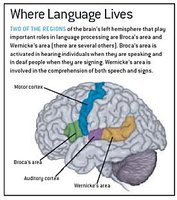
Language processing involves specific brain regions:
Broca’s area: Helps with speaking languages
Wernicke’s area: Helps with understanding languages
Memory
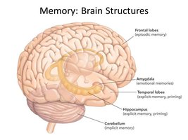
Memory involves storage and retrieval of information, with several types:
Declarative Memory: Facts; includes short-term (limited) and long-term (limitless, declines with age)
Procedural Memory: Skills
Motor Memory: Motor skills
Emotional Memory: Emotional responses
Factors influencing memory: Emotional state, rehearsal, association, automatic memory (formed via first impression)
Brain Wave Patterns
Continuous electrical activity in the brain is measured by an electroencephalogram (EEG), producing wave-like patterns:
Alpha (8-13 Hz): Regular, rhythmic; calm wakefulness
Beta (14-30 Hz): Less regular; mentally alert and concentrating
Theta (4-7 Hz): Irregular; most common in children, appear when concentrating
Delta (<5 Hz): High amplitude; observed during sleep, in awake adults indicates brain damage
Sleep/Wake Cycles
Sleep is divided into non-rapid eye movement (NREM) and rapid eye movement (REM) stages:
NREM: Four stages, from relaxation (alpha waves) to deep sleep (delta waves)
REM: Muscles inhibited except eyes and diaphragm; dreaming occurs
Regulation: Circadian rhythm controlled by the hypothalamus and hormones.
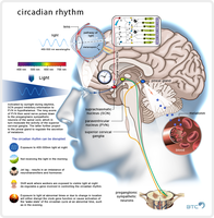
Importance: Hypotheses include memory and emotional analysis, elimination of unneeded synapses.
Consciousness
Consciousness is undefined but involves simultaneous activity of large areas of the cerebral cortex. It is holistic and interconnected, measured on a gradient from alertness to coma.
Brain Protection
Protective Structures
The brain is protected by skull bones, cranial meninges, cerebrospinal fluid (CSF), and the blood-brain barrier.
Cranial Meninges: Three protective membranes of dense, irregular connective tissue
Cerebrospinal Fluid: Fluid with same density as brain, fills ventricles and surrounds brain; provides buoyancy, temperature regulation, and waste removal
Blood Brain Barrier: Separates CSF and brain extracellular fluid from blood; blocks toxins and pathogens
Meninges
Meninges are connective tissue membranes that cover and protect the CNS, protect blood vessels, enclose the venous system, contain CSF, and partition the skull.
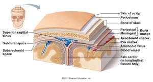
Dura Mater
The dura mater is the strongest meninx, a bi-layered sheet:
Periosteal layer: Attaches to skull
Meningeal layer: External covering of the brain
Separations form dural venous sinuses and partitions such as the falx cerebri, falx cerebelli, and tentorium cerebelli.
Arachnoid Mater
The arachnoid mater is the middle meninx, separated from the dura mater by the subarachnoid space filled with CSF.
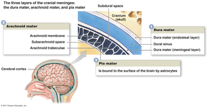
Pia Mater
The pia mater is the innermost meninx, embedded with vasculature and bound to the surface of the brain by astrocytes.
Cerebrospinal Fluid (CSF)
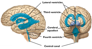
CSF is formed in choroid plexuses of ventricles where blood vessels contact ependymal cells, extracting and converting plasma to CSF. It circulates through ventricles, subarachnoid space, and central canal, and is reabsorbed into the bloodstream via dural sinuses (arachnoid granules).
Properties: Similar in composition to blood plasma, but with more Na+, Cl-, and H+, and less Ca2+ and K+. About 500 ml produced daily.
Blood Brain Barrier (BBB)
The blood brain barrier protects the brain by blocking toxins, pathogens, and large molecules from entering. It allows selective transport of essential nutrients and maintains homeostasis for neurons.
What Can Cross the BBB? Small, lipid-soluble molecules (e.g., oxygen, carbon dioxide), specific nutrients via transport systems (e.g., glucose via GLUT1 transporter), water through aquaporins.
Structure | Function |
|---|---|
Dura Mater | Strongest, protects brain, forms partitions and sinuses |
Arachnoid Mater | Middle layer, contains CSF, provides cushioning |
Pia Mater | Innermost, follows brain contours, contains blood vessels |
CSF | Buoyancy, protection, waste removal, homeostasis |
Blood Brain Barrier | Selective permeability, protection from toxins |
Example: The BBB prevents most drugs and pathogens from entering the brain, but allows oxygen and glucose to pass, ensuring proper neuronal function.
