 Back
BackCentral Nervous System (CNS) and Its Protection: Key Concepts and Structures
Study Guide - Smart Notes
 Tailored notes based on your materials, expanded with key definitions, examples, and context.
Q1. What, anatomically, is included in the CNS?
Background
Topic: Central Nervous System Anatomy
This question tests your understanding of the basic anatomical components that make up the central nervous system (CNS), which is a foundational concept in neuroanatomy.
Key Terms:
Central Nervous System (CNS): The part of the nervous system consisting of the brain and spinal cord.
Brain: The organ located within the skull, responsible for integrating sensory information and coordinating body functions.
Spinal Cord: The long, thin, tubular structure running from the brainstem down the vertebral column, transmitting signals between the brain and the rest of the body.
Step-by-Step Guidance
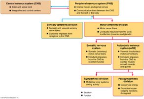
Recall that the nervous system is divided into the central nervous system (CNS) and the peripheral nervous system (PNS).
Identify the two main anatomical structures that make up the CNS: the brain and the spinal cord.
Consider the location of these structures: the brain is housed within the skull, and the spinal cord is protected by the vertebral column.
Try solving on your own before revealing the answer!
Q2. How would you summarize the function of the CNS?
Background
Topic: CNS Function
This question is about the overall role of the CNS in processing sensory input and generating motor output, which is central to understanding how the nervous system controls the body.
Key Terms:
Sensory Input: Information received from sensory receptors about internal and external changes.
Integration: The CNS processes and interprets sensory input and decides what should be done at each moment.
Motor Output: The CNS sends signals to muscles and glands to elicit a response.
Step-by-Step Guidance
Think about the flow of information: sensory information comes into the CNS from the body via the PNS.
The CNS integrates and interprets this information to make decisions.
Motor commands are then sent out from the CNS to the body to produce a response.
Try solving on your own before revealing the answer!
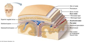
Q1. Label the spinal meninges.
Background
Topic: Protection of the CNS – Meninges
This question focuses on the three protective membranes (meninges) that surround the brain and spinal cord, which are essential for CNS protection.
Key Terms:
Dura Mater: The tough, outermost membrane.
Arachnoid Mater: The middle, web-like membrane.
Pia Mater: The delicate, innermost membrane that adheres to the surface of the CNS.
Step-by-Step Guidance
Recall the order of the meninges from outermost to innermost: dura mater, arachnoid mater, pia mater.
Identify these layers in a cross-sectional diagram of the spinal cord or brain.
Note that the same names are used for both cranial and spinal meninges.
Try solving on your own before revealing the answer!
Q2. What do these names mean?
Background
Topic: Etymology of Meningeal Layers
This question asks you to consider the literal meanings of the names of the meningeal layers, which often reflect their structure or appearance.
Key Terms:
Dura Mater: "Tough mother" (Latin), indicating its durable nature.
Arachnoid Mater: "Spider-like mother," referring to its web-like appearance.
Pia Mater: "Gentle mother," describing its delicate structure.
Step-by-Step Guidance
Break down each term into its Latin roots to understand the meaning.
Relate the meaning to the physical characteristics of each layer.
Try solving on your own before revealing the answer!
Q1. What are the names of the ventricles?
Background
Topic: Ventricular System of the Brain
This question is about the fluid-filled spaces within the brain that produce and circulate cerebrospinal fluid (CSF).
Key Terms:
Lateral Ventricles: Paired structures, one in each cerebral hemisphere.
Third Ventricle: Located in the midline, between the two halves of the thalamus.
Fourth Ventricle: Located between the brainstem and the cerebellum.
Step-by-Step Guidance
Recall the sequence and location of the ventricles in the brain.
Identify each ventricle in a diagram of the brain.

Try solving on your own before revealing the answer!
Q2. What part of the spinal cord contains most of the CSF? (Does the spinal cord have ventricles?)
Background
Topic: CSF Circulation in the CNS
This question explores where cerebrospinal fluid is found in the spinal cord and whether the spinal cord contains ventricles like the brain.
Key Terms:
Central Canal: The small channel running through the center of the spinal cord, containing CSF.
Subarachnoid Space: The space between the arachnoid mater and pia mater, filled with CSF.
Step-by-Step Guidance
Recall that the brain has ventricles, but the spinal cord does not have large ventricles.
Identify the central canal and subarachnoid space as the main locations for CSF in the spinal cord.
Try solving on your own before revealing the answer!
