 Back
BackCentral Nervous System: Structure and Function of the Brain
Study Guide - Smart Notes
 Tailored notes based on your materials, expanded with key definitions, examples, and context.
The Embryologic Perspective
Development of the Neural Tube
The central nervous system (CNS) originates from the neural tube during embryonic development. The anterior end of the neural tube forms three primary brain vesicles, which further differentiate into five secondary vesicles, giving rise to the major regions of the adult brain.
Primary Vesicles: Prosencephalon, Mesencephalon, Rhombencephalon
Secondary Vesicles: Telencephalon (cerebrum), Diencephalon (thalamus & hypothalamus), Mesencephalon (midbrain), Metencephalon (pons & cerebellum), Myelencephalon (medulla oblongata)
The brainstem consists of the midbrain, pons, and medulla oblongata.
The spinal cord remains tube-shaped, with posterior neurons for sensory functions and anterior neurons for motor functions.
The ventricular system of the adult brain is derived from the hollow neural tube, and cerebrospinal fluid (CSF) circulates through these ventricles.
The Central Nervous System: Major Regions of the Brain
Overview of Brain Regions
The adult brain is divided into four main regions, each with distinct functions and anatomical features.
Cerebrum: Largest part, divided into lobes and hemispheres, responsible for higher cognitive functions.
Cerebellum: Second largest, coordinates movement and posture.
Diencephalon: Contains structures ending in 'thalamus,' involved in sensory relay and homeostasis.
Brain Stem: Oldest region, controls homeostatic and autonomic functions.
Key Anatomical Terms
Lobe: Brain section named for the overlying cranial bone.
Gyrus: Ridge of a brain fold.
Sulcus: Groove between gyri; major sulci separate lobes.
Fissure: Deep groove, such as the longitudinal fissure separating hemispheres.
Cortex: Thin outer layer of gray matter.
Nucleus: Cluster of gray matter deep within white matter.
Prominent Landmarks of the Cerebrum
Central Sulcus: Separates frontal and parietal lobes.
Lateral Sulcus: Separates temporal lobe from frontal and parietal lobes.
Longitudinal Fissure: Divides right and left hemispheres.
Precentral Gyrus: Primary motor cortex, anterior to central sulcus.
Postcentral Gyrus: Primary somatosensory cortex, posterior to central sulcus.
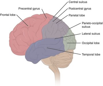
Lobes of the Cerebrum
Frontal Lobe: Motor functions, short-term memory, decision making, personality, Broca’s area (language production).
Parietal Lobe: General sensation, taste.
Temporal Lobe: Auditory sensation, long-term memory, Wernicke’s area (language comprehension).
Occipital Lobe: Visual perception.
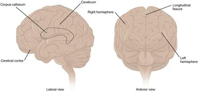
Hemispheres and Connections
The cerebrum is divided into left and right hemispheres by the longitudinal fissure.
The corpus callosum connects the two hemispheres, allowing communication between them.
The cerebral cortex is the outermost layer, responsible for higher-order functions.
Basal nuclei are involved in cognitive processing and motor planning.

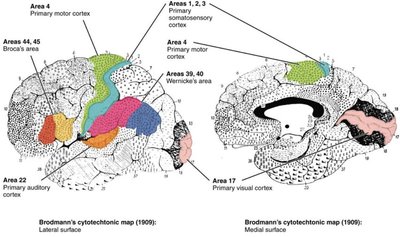
Functional Areas of the Cerebral Cortex
Brodmann’s Areas
Brodmann’s areas are regions of the cerebral cortex defined by their cytoarchitecture, each associated with specific functions.
Broca’s Area: Language production (left hemisphere).
Prefrontal Cortex: Personality, short-term memory, consciousness.
Premotor Area: Planning of movement.
Frontal Eye Fields: Eye movement control.
Subcortical Structures of the Cerebrum
Basal Nuclei
The basal nuclei are deep gray matter structures that play a crucial role in movement control, learning, and memory.
Components: Caudate, Putamen, Globus pallidus (collectively called the striatum).
Functions: Movement regulation, cognitive processing, and motor planning.
Basal Nuclei Pathways
Direct Pathway: Disinhibits the thalamus, facilitating movement (activated by dopamine).
Indirect Pathway: Reinforces inhibition of the thalamus, suppressing movement (inhibited by dopamine).

Other Subcortical Structures
Basal Forebrain: Involved in learning and memory, produces acetylcholine (ACh).
Limbic Cortex: Regulates emotion, memory, and behavior.
Hippocampus: Essential for memory formation.
Amygdala: Involved in emotion and memory, especially fear and aggression.
The Diencephalon
Major Components
Thalamus: Relay center for all sensory information except olfaction; processes and transmits signals to the cerebral cortex.
Hypothalamus: Maintains homeostasis, regulates the autonomic nervous system and endocrine system, involved in memory and emotion, senses blue light for circadian rhythms.
Epithalamus: Contains the pineal gland, which secretes melatonin for sleep regulation.

The Brain Stem
Structure and Function
Midbrain: Coordinates sensory information (visual, auditory, somatosensory), contains cerebral peduncles, tectum, and tegmentum, produces dopamine for basal nuclei, contains cranial nerves III and IV.
Pons: Main connection between cerebellum and brain, important for REM sleep, contains cranial nerves V–VIII, forms the floor of the fourth ventricle.
Medulla Oblongata: Regulates breathing, heart rate, and blood vessel function, contains pyramids where corticospinal tracts decussate, contains cranial nerves IX–XII, suppressed by depressant drugs.

The Cerebellum
Structure and Function
The cerebellum, or "little brain," is responsible for coordinating movement and maintaining posture. It is separated into hemispheres by the vermis and contains anterior and posterior lobes. The external gray matter is called folia, and the inner white matter is known as the arbor vitae.
Coordinates voluntary movements and balance.
Can modify motor commands to ensure smooth execution of movement.

Functional Systems of the Brain
Limbic System
Composed of the amygdala, basal nuclei, fornix, mammillary body, cingulate gyrus, hippocampus, and hypothalamus.
Responsible for emotion, memory, and behavior.
Strong connection between olfaction (smell) and memory.
The amygdala is involved in emotional responses, including the "four F's": fighting, fleeing, feeding, and reproduction.
Reticular Formation
Network of neurons extending from the medulla to the cortex.
Keeps the cortex alert and regulates consciousness.
Filters sensory information and is inhibited by alcohol and sleep-inducing drugs.
Disorders such as ADHD can affect this system, impacting focus and attention.
Review Questions
What are the four lobes of the cerebrum?
What is the function of the precentral and postcentral gyri?
What is the diencephalon responsible for?
What are the components of the brain stem?
