 Back
Central Nervous System: Structure and Function
Study Guide - Smart Notes
 Tailored notes based on your materials, expanded with key definitions, examples, and context.
Central Nervous System Overview
Definition and Components
The central nervous system (CNS) consists of the brain and spinal cord. It is responsible for integrating sensory information, coordinating motor output, and supporting higher functions such as intellect, behavior, and personality.
Brain: The control center for behavior, intellect, and personality, composed of billions of neurons.
Spinal Cord: Provides sensory and motor innervation below the head, serves as a pathway for signals, and is the integration center for reflexes.
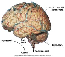
Directional Terminology
Directional terms are used to describe locations within the CNS:
Rostral: Toward the nose/forehead (anterior)
Caudal: Toward the tail/spinal cord (posterior)
Gray and White Matter in the CNS
Brain and Spinal Cord Organization
The CNS contains two distinct types of tissue:
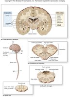
Gray Matter: Composed of neuronal cell bodies, dendrites, and unmyelinated axons. In the brain, it forms the cortex (outer layer) and deep nuclei (inner clusters).
White Matter: Consists mainly of myelinated axons, responsible for rapid communication between regions.
Basic Organization of the Brain
Major Regions
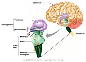
The brain is divided into four main regions:
Brain Stem: Controls basic life functions and connects the brain to the spinal cord.
Cerebellum: Coordinates movement and balance.
Diencephalon: Contains thalamus, hypothalamus, and epithalamus; central core of the forebrain.
Cerebrum: Largest part, responsible for higher functions.
Cerebrum
Structure and Surface Features
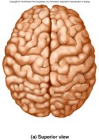
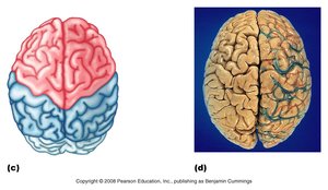
The cerebrum is the most rostral part of the brain, covering the diencephalon and brain stem. It is divided into two hemispheres and makes up about 80% of brain mass.
Gyri: Elevated ridges on the surface.
Sulci: Shallow grooves between gyri.
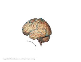
Fissures: Deep grooves separating major regions (e.g., longitudinal fissure between hemispheres, transverse fissure between cerebrum and cerebellum).
Cerebral Lobes
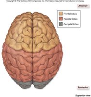
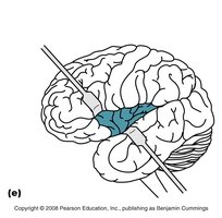
The cerebrum is divided into five lobes, named for the overlying skull bones:
Frontal Lobe: Motor functions, reasoning, planning.
Parietal Lobe: Somatosensory perception.
Occipital Lobe: Visual processing.
Temporal Lobe: Auditory processing.
Insula: Taste and balance, covered by other lobes.
Cerebral Cortex
The cerebral cortex is a thin layer (2-4 mm) of gray matter responsible for consciousness, motor initiation, communication, memory, and comprehension.
Functional Areas:
Sensory Areas: Perception of sensations (e.g., somatosensory, visual, auditory, gustatory).
Association Areas: Higher processing and integration of sensory information.
Motor Areas: Control voluntary movements.
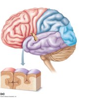
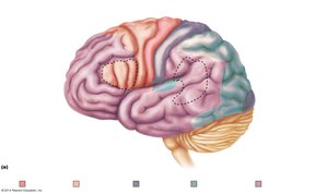
Functional Areas of the Cerebral Cortex
Sensory Areas:
Parietal lobe: Primary somatosensory cortex (touch, pressure, temperature)
Occipital lobe: Primary visual cortex
Temporal lobe: Primary auditory cortex
Insula: Gustatory cortex (taste) and balance
Association Areas:
Sensory association areas: Higher processing of specific senses
Multimodal association areas: Integrate information from multiple senses
Motor Areas:
Primary motor cortex: Initiates voluntary movement (precentral gyrus)
Premotor cortex: Coordinates complex movements
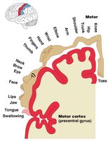
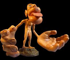
Homunculus Maps
The motor and sensory homunculus are maps on the cortex showing which regions control or perceive specific body parts. Larger representations indicate greater sensitivity or motor control. 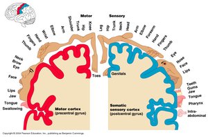
Cerebrum: White Matter and Nuclei
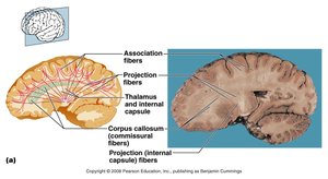
White Matter
Corpus Callosum: Major tract connecting the two hemispheres, allowing communication.
Other tracts: Association, projection, and commissural fibers.
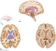
Basal Nuclei
Basal nuclei (ganglia): Deep clusters of gray matter involved in motor control, working with the motor cortex.
Dysfunction (e.g., Parkinson's disease) leads to motor deficits.
Diencephalon
Components and Functions
The diencephalon forms the central core of the forebrain and includes:
Thalamus: Relay station for sensory information; processes and sends signals to the cortex.
Hypothalamus: Controls autonomic functions, endocrine system, body temperature, hunger, thirst, and circadian rhythms; produces hormones and interacts with the pituitary gland.
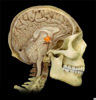
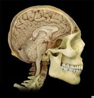
Epithalamus: Contains the pineal gland, which secretes melatonin to regulate sleep.
Brain Stem
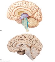
Structure and Divisions
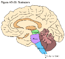
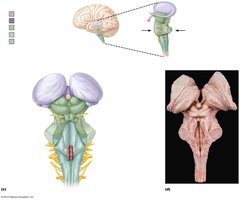
The brain stem is the most caudal region and consists of:
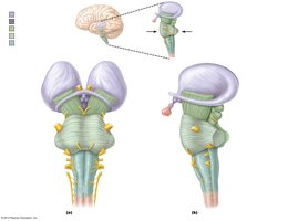
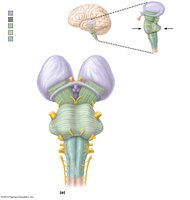
Midbrain: Contains cerebral peduncles, superior and inferior colliculi (visual and auditory reflexes).
Pons: Bridge between brainstem and cerebellum; involved in movement coordination.
Medulla Oblongata: Contains autonomic nuclei for vital functions (respiratory, cardiac, vasomotor centers).
Cerebellum
Structure and Function
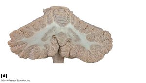
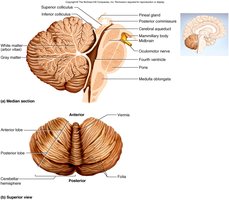
The cerebellum is located dorsal to the pons and medulla, consisting of two hemispheres connected by the vermis. It coordinates body movements, maintains posture and balance, and stores motor memories.
Receives information from the motor cortex and proprioceptors.
Compares planned movement to actual movement and sends corrective signals.
Ventricles of the Brain
Structure and Function
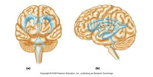
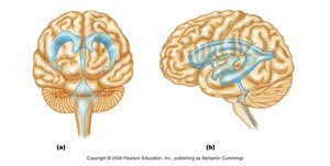
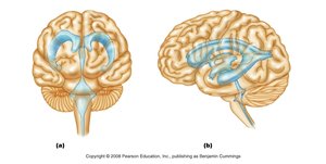
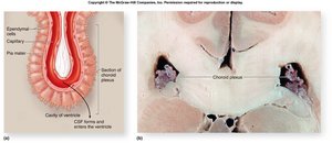
The ventricles are cavities filled with cerebrospinal fluid (CSF), lined with ependymal cells, and continuous with the central canal of the spinal cord.
Lateral ventricles: Located in cerebral hemispheres, separated by septum pellucidum.
Third ventricle: Located in diencephalon, connected to lateral ventricles by interventricular foramen.
Fourth ventricle: Located in brainstem, connects to central canal and subarachnoid space.
Cerebral aqueduct: Connects third and fourth ventricles.
Protection of the Brain
Meninges
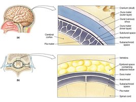
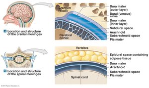
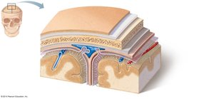
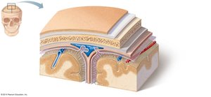
The meninges are three protective layers surrounding the brain:
Dura Mater: Tough, outermost layer with periosteal and meningeal sublayers; forms dural sinuses for venous blood return.
Arachnoid Mater: Middle layer with subarachnoid space (contains CSF) and arachnoid villi (CSF absorption).
Pia Mater: Delicate, innermost layer, rich in blood vessels, adheres to brain surface.
Cerebrospinal Fluid (CSF)
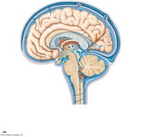
CSF cushions the brain and spinal cord, provides nutrients, removes waste, and carries chemical signals.
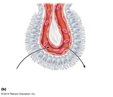
Produced by the choroid plexus in each ventricle; flows through ventricles, subarachnoid space, and is absorbed into dural sinuses via arachnoid granulations.
Blood Brain Barrier (BBB)
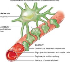
The blood brain barrier protects the brain from toxins and pathogens by restricting passage of substances from the blood.
Formed by tight junctions between endothelial cells, supported by astrocytes.
Allows lipid-soluble molecules (e.g., alcohol, nicotine) to pass.
Spinal Cord
Structure and Function
The spinal cord extends from the foramen magnum to the first or second lumbar vertebra, divided into 31 segments with paired spinal nerves.
Responsible for sensory and motor innervation below the head.
Provides a two-way conduction pathway and integrates reflexes.
Gray and White Matter in the Spinal Cord
Gray Matter: Centrally located, H-shaped, contains neuron cell bodies, dendrites, and neuroglia.
White Matter: Surrounds gray matter, contains myelinated and unmyelinated axons.
Spinal Cord Pathways
Ascending Tracts: Carry sensory information to the brain.
Descending Tracts: Carry motor commands from the brain to the body.
Gray Matter Regions
Gray Commissure: Crossbar of the H, surrounds central canal.
Dorsal Horn: Receives sensory information; divided into somatic and visceral regions.
Ventral Horn: Contains motor neuron bodies; divided into somatic (skeletal muscle) and visceral (smooth/cardiac muscle, glands) regions.
Protection of the Spinal Cord
Protective Structures
The spinal cord is protected by the vertebral column, meninges, and cerebrospinal fluid.
Meninges are similar to those in the brain, but the dura mater is separated from the vertebrae by the epidural space (contains fat).
Communication Between CNS and Body
Ascending and Descending Pathways
Ascending Pathways: Sensory information travels from peripheral receptors to the brain via multi-neuronal pathways.
Descending Pathways: Motor output travels from the motor cortex to the spinal cord.
Summary Table: Major CNS Structures and Functions
Structure | Main Function |
|---|---|
Brain | Integration, higher functions |
Spinal Cord | Conduction, reflexes |
Cerebrum | Consciousness, motor/sensory processing |
Diencephalon | Sensory relay, homeostasis |
Brain Stem | Basic life functions, cranial nerve attachment |
Cerebellum | Coordination, balance |
Meninges | Protection |
CSF | Cushioning, nutrient/waste transport |
Blood Brain Barrier | Protection from toxins |
