 Back
Central Nervous System: Structure and Functional Organization
Study Guide - Smart Notes
 Tailored notes based on your materials, expanded with key definitions, examples, and context.
Central Nervous System Overview
Major Components of the CNS
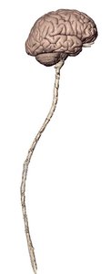
The central nervous system (CNS) consists of the brain and spinal cord. These structures are responsible for integrating sensory information and coordinating bodily functions.
Brain: The control center for processing information, thought, and emotion.
Spinal Cord: The main pathway for transmitting signals between the brain and the rest of the body.
Protection of the CNS
The CNS is protected by several structures:
Meninges: Three connective tissue layers (dura mater, arachnoid mater, pia mater) that surround the brain and spinal cord.
Ventricles: Internal cavities within the brain filled with cerebrospinal fluid (CSF).
Cerebrospinal Fluid (CSF): Cushions the CNS and removes waste products.
Directional Terms in CNS Anatomy
Unique Directional Terms
Directional terms are used to describe locations within the CNS:
Rostral: Toward the nose or higher brain regions (anterior/superior).
Caudal: Toward the tail or lower brain regions (posterior/inferior).
Major Regions of the Brain
Four Main Regions
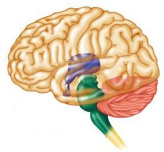
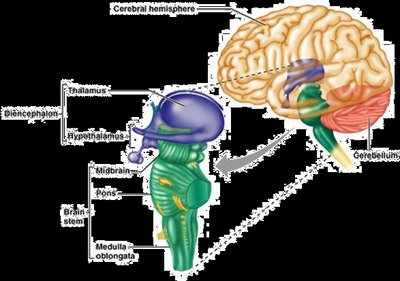
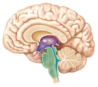
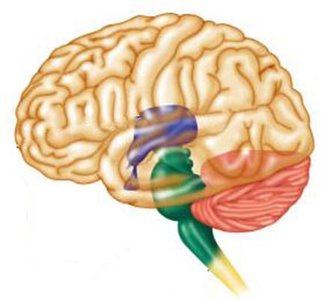
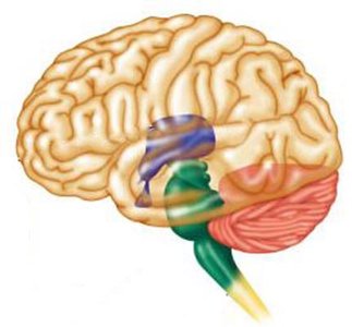
The adult brain is divided into four main regions, each with distinct functions:
Cerebrum: Largest region, responsible for higher cognitive functions.
Diencephalon: Includes the thalamus and hypothalamus, involved in sensory relay and homeostasis.
Brainstem: Controls basic life functions; includes the midbrain, pons, and medulla oblongata.
Cerebellum: Coordinates movement and balance.
The Cerebrum
Basic Organization
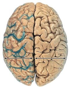
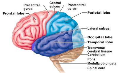
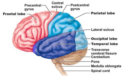
The cerebrum is the largest part of the brain, comprising about 83% of its mass. It consists of two hemispheres and is characterized by surface landmarks:
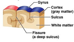
Fissures: Deep grooves separating major regions.
Sulci: Shallow grooves on the surface.
Gyri: Ridges between sulci.
Internal Structure
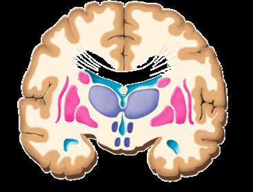
The cerebrum is organized into three main types of tissue:
Cerebral Cortex: Outer layer of gray matter responsible for conscious thought.
Basal Ganglia: Deep gray matter involved in movement regulation.
White Matter: Myelinated axons connecting different brain regions.
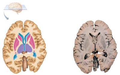
Surface Landmarks of the Cerebrum
Definitions and Examples
Surface landmarks are important for identifying regions and functions:
Sulci: Grooves on the surface of the cerebrum.
Fissures: Very deep grooves separating major regions.
Gyri: Ridges between sulci.
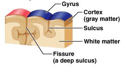
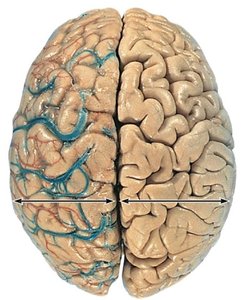
Major Fissures
Longitudinal Fissure: Separates right and left cerebral hemispheres.
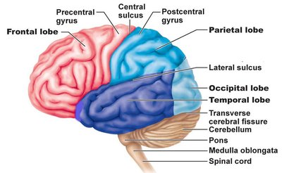
Transverse Fissure: Separates cerebrum from cerebellum.
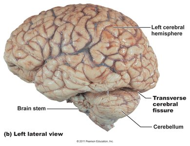
Major Sulci and Gyri
Central Sulcus: Separates frontal and parietal lobes.
Lateral Sulcus: Separates temporal lobe from frontal and parietal lobes.
Parieto-Occipital Sulcus: On the medial surface, separates parietal and occipital lobes.
Precentral Gyrus: Primary motor cortex.
Postcentral Gyrus: Primary somatosensory cortex.
Cerebral Lobes
Identification and Location
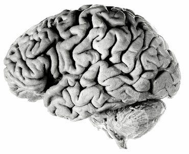
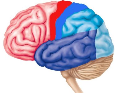
The cerebrum is divided into five lobes, each named for the overlying skull bones:
Frontal Lobe: Anterior to central sulcus, superior to lateral sulcus.
Parietal Lobe: Posterior to central sulcus, superior to lateral sulcus, anterior to parieto-occipital sulcus.
Temporal Lobe: Inferior to lateral sulcus, anterior to occipital lobe.
Occipital Lobe: Posterior to parieto-occipital sulcus and temporal lobe.
Insula: Deep within lateral sulcus, covered by parts of temporal, parietal, and frontal lobes.
Medial Surface Landmarks
Parieto-Occipital Sulcus: Separates occipital lobe from parietal lobe on the medial surface.
Insula
The insula is considered the "fifth lobe" and is located deep within the lateral sulcus.
Summary Table: Surface Landmarks and Lobes
Lobe | Location | Key Landmark |
|---|---|---|
Frontal | Anterior to central sulcus, superior to lateral sulcus | Precentral gyrus |
Parietal | Posterior to central sulcus, superior to lateral sulcus, anterior to parieto-occipital sulcus | Postcentral gyrus |
Temporal | Inferior to lateral sulcus, anterior to occipital lobe | Lateral sulcus |
Occipital | Posterior to parieto-occipital sulcus and temporal lobe | Parieto-occipital sulcus |
Insula | Deep within lateral sulcus | Covered by other lobes |
Functional Areas of the Cerebral Cortex
Overview of Functional Areas
The cerebral cortex is the site of conscious mind and is divided into three main functional areas:
Sensory Areas: Process sensory information and provide conscious awareness of sensation.
Motor Areas: Initiate and control voluntary movements.
Association Areas: Integrate information to enable complex and purposeful action.
All neurons in the cerebral cortex are interneurons.
Motor Areas
Primary Motor Cortex
Premotor Cortex
Frontal Eye Field
Broca’s Area
Multimodal Association Areas
Posterior Association Area
Wernicke’s Area
Anterior Association Area (Prefrontal Cortex)
Limbic Association Area
Sensory Areas
Somatosensory Areas
Visual Areas
Auditory Areas
Gustatory Cortex
Olfactory Cortex
Vestibular Cortex
Additional info:
The organization and landmarks of the cerebrum are fundamental for understanding brain function and clinical neuroanatomy.
Functional areas are mapped using techniques such as MRI and histological staining.
